The most common type of heart disease in cats is cardiomyopathy, the most common of which is hypertrophic cardiomyopathy HCM (accounting for about 60% of all cases), others such as restrictive cardiomyopathy RCM, non-specific myocardial disease NCM (approximately 20-30%) and dilated cardiomyopathy DCM ARVC may occur but the probability is low.
💗Before there are clinical symptoms of cardiomyopathy in cats, abnormalities can sometimes be detected through auscultation, but a large part of them may not be detected through general auscultation and other physical examinations.
Cats that do not yet have clinical symptoms usually need to undergo physical examination, chest xray, and cardiac biomarker (such as proBNP) tests for detection. However, cats with heart problems may not necessarily detect abnormalities by auscultation, and xray is also difficult to detect. Only the outline of the heart cannot be used to know the internal conditions. In addition, cardiac biomarkers in the blood cannot be completely detected when used to detect heart disease in asymptomatic cats. Therefore, if further confirmation of diagnosis is required, cardiac ultrasound examination is still required.
According to statistics in human medicine, about 1/200-1/500 people will have hypertrophic cardiomyopathy. However, studies in cats have found that about one in seven cats will have myocardium detected under cardiac ultrasound. Hypertrophic condition. Therefore, regular physical examination during the asymptomatic period is still very important.
💔The most common symptoms of cats with heart failure are rapid breathing, paralysis due to arterial thrombosis, and even sudden death.
🙋What kind of cats are at high risk of heart disease? Are cardiac ultrasound examinations recommended?
👩⚕️
➡️Medical history appears
◦ Fainting or symptoms similar to epilepsy but without other neurological symptoms
◦ Weakness, exercise intolerance, open breathing
◦ People with endocrine diseases
◦ People who are heartworm positive
◦ Cat breeds with heart disease gene MyBPC3 mutations
◦ People with unknown fever origin
➡️Or physical examination findings
◦ Heart murmur
◦ Galloping sound or systolic click (systolic click)
◦ Irregular heartbeat
◦ Ascites
◦ Sudden paralysis, etc.
◦ Jugular venous distention
◦ Weak femoral artery, etc.
◦ Or cats older than nine years old need some treatment, which may easily lead to heart failure, etc.
◦ General anesthesia
◦ Infusion therapy
◦ Long-acting steroids or long-term use of steroids are required.
🙋Common types of heart disease in cats
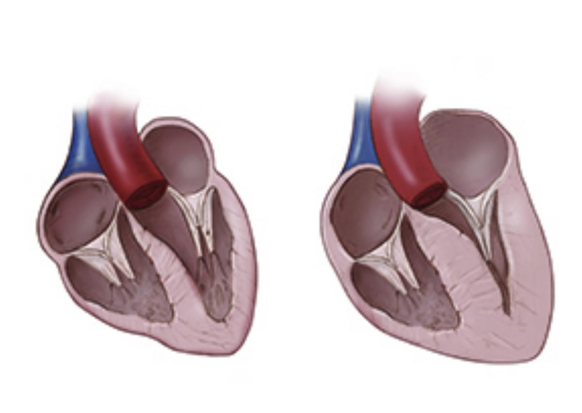
👩⚕️Hypertrophic cariomyopathy
➡️Definition: When there is concentric hypertrophy of the myocardium, and other systemic diseases such as hypertension and hyperthyroidism, and other structural changes such as aortic stenosis are excluded, it is called hypertrophic cardiomyopathy.
➡️The cause of the occurrence is unknown. In addition to the genetic mutation related between Maine Coon cats and Ragdoll cats, other purebred cats such as American shorthair cats, British shorthair cats, Persian cats and leopard cats may have related genetic genes, but currently there is no such genetic mutation. complete research
😺There is also a type of temporary myocardial thickening that has a better prognosis
‣ Usually younger
‣ It is usually suspected that the myocardium is rapidly damaged due to anesthesia, trauma or the use of steroids, resulting in edema, etc. Myocardial hypertrophy may occur temporarily and may return to normal after 2-5 months.
‣ Compared with ordinary hypertrophic cardiomyopathy, the myocardium is usually not very thick, and the atrium is not as large as that of ordinary hypertrophic cardiomyopathy.
➡️Diagnosis: Cardiac hypertrophy will appear under cardiac ultrasound. Severe cases are easy to diagnose, but mild cases are more challenging to diagnose because they may be partially hypertrophic. You also need to refer to the cat’s body shape to judge.The size of the left atrium is also related to the severity of the disease. Generally, the larger the left atrium, the higher the chance of pulmonary hydrops and thrombosis. Because the heart is a 3D structure, some cats may have severe atrial dilation, but some cats may have severe atrial dilation. The dilation of the left atrial appendage is relatively serious, so both objective measurement and subjective judgment need to be referenced together.
👩⚕️Dilated cardiomyopathy
➡️Definition: Left ventricular dilation, myocardial contraction deterioration and exclusion of systemic hypertension, membrane disease and ischemic disease, etc.
➡️Cause of occurrence: unknown. Before 1987, the lack of taurine was the main reason. But now commercial feeds do not have such problems. However, some families use fresh food cooked by themselves or feed their cats. Vegetarian feeds, etc. may cause taurine deficiency.
➡️Diagnosis: Under cardiac ultrasound, it will be seen that the diameter of the left ventricle at the end of systole becomes larger. In order to compensate for the insufficient contractility, the myocardium will initially form centrifugal hypertrophy. The so-called dilation refers to the myocardial expansion after the myocardial compensation. reaction, the real cause of the disease is abnormal contraction ability
😿It is usually difficult to detect heart failure before symptoms occur. When heart failure occurs, the contractile capacity may already be very poor (FS<15%)
👩⚕️Restrictive cardiomyopathy
➡️Definition: Abnormal diastolic ability of the left heart, divided into endocardial, subendocardial and myocardial fibrosis. Like the myocardium of hypertrophic cardiomyopathy, myocardial fibrosis can also occur. , but the difference is that the myocardium will become thicker in hypertrophic cardiomyopathy, but not in restrictive cardiomyopathy, and fibrosis may be more severe in restrictive cardiomyopathy. When the myocardium hardens, it loses its elasticity, just like a balloon originally made of latex can be easily inflated, but a balloon made of rubber requires more force to inflate, which will increase the left heart pressure throughout the end-diastole, thus When the pressure in the pulmonary capillaries increases and more water enters the space between capillaries and tissues than the lymphatic system’s ability to remove water, pulmonary hydrops or pleural effusion will occur. Although the right atrium will also become enlarged, ascites is relatively rare in cats. .
➡️Cause of occurrence: unknown, may be related to genes or infection.
Restrictive cardiomyopathy, left ventricular endocardium thickening, fibrosis and whitening, left atrial appendage dilation
➡️Diagnosis: Difficult to diagnose before the asymptomatic period and before the atria become enlarged
‣ Under ultrasound, you will see a left heart with normal contractility and normal structure, as well as a dilated left atrium and right atrium, etc.
‣ Diagnosis requires abnormal diastolic capacity and exclusion of hypertrophic cardiomyopathy
‣ Under cardiac ultrasound, if the heartbeat is <180bpm, a high E wave can be seen, and a reduced E' wave can be seen on tissue duplex, and in some cases of endocardial fibrosis, cardiac ultrasound can detect in specific areas A more reverberant image will appear.
‣ In chest xrays with restrictive cardiomyopathy, the proportion of pleural effusion is higher than that of pulmonary effusion.
👩⚕️Arrhythmogenic right ventricular cardiomyopathy ARVC
➡️Definition: Pathologically similar to human ARVC, you will see thinning of the right ventricular free wall (may be diffuse or local thinning). These thinning areas are normal myocardial fibers or It is replaced by fibrofatty tissue. Under dissection, it can be seen that the free wall becomes thinner and transparent, and the right atrium becomes severely enlarged. This disease is relatively rare in cats.
➡️Cause of occurrence: unknown. It is usually a hereditary disease in humans but there is currently no relevant genetic research on cats.
Arrhythmogenic cardiomyopathy (ARVC) enlargement and thinning of the right heart
➡️Symptoms: Usually when it is discovered, symptoms of right heart failure will appear, such as hepatic venous enlargement, ascites, etc., and arrhythmias such as premature ventricular contraction, ventricular tachycardia, etc. may also occur.
➡️Diagnosis: Under cardiac ultrasound, there will be enlargement of the right ventricle and atrium, tricuspid valve turbulence, and local lack of mobility of the right ventricular free wall. It needs to be distinguished from tricuspid valve hypoplasia. Although the difference between the two is difficult, overall It is said that the right ventricular wall mobility of ARVC is relatively poor, and the degree of tricuspid valve opening and closing will be smaller than that of normal cats. However, in cases of tricuspid valve hypoplasia, the right ventricular wall mobility will be greater than that of normal cats, and the opening and closing of the tricuspid valve will be smaller than that of normal cats. The cat will also be larger than a normal cat
➡️Prognosis: Overall prognosis is poor
◦ Treatment: If ventricular tachycardia occurs, which may lead to sudden death of the cat, arrhythmia drugs can be given
👩⚕️In addition to the above-mentioned heart diseases, there are also
➡️Compacting cardiomyopathy left ventricular noncompaction
➡️Nonspecific phenotype of heart disease
➡️Myocardial hypertrophy caused by hyperthyroidism: Some cats with hyperthyroidism will cause concentric left ventricular hypertrophy due to stimulation of the thyroid gland, but usually the increased thickness due to hyperthyroidism does not exceed 2mm, so if the end-diastolic left ventricular thickness of hyperthyroid cats is found It is already larger than 7mm. Most of these cats already have hypertrophic cardiomyopathy, but hyperthyroidism may make the myocardium more hypertrophic.
🙋With so many types of heart disease, how to stage them?
👩⚕️Although there are many types of diseases, fortunately the staging rules are similar, and the prognosis and treatment of different stages are also different.
💟A Cat breeds that are more susceptible to heart disease, such as Maine Coons, but there is no evidence of cardiomyopathy
💟B1: There are no clinical symptoms yet, but cardiac ultrasound shows cardiomyopathy, but there is no obvious dilation of the left and right atria. Cats at this stage have a low chance of developing symptoms of heart failure and blood clots. The probability of death within one year is very low <10%. No drug treatment is required at this stage, but regular cardiac ultrasound tracking is still required within six months to one year. Current research shows that left ventricular outlet dynamic obstruction is required, and drug treatment and follow-up are required. There is not much difference in the five-year survival rate without drug treatment, so administration is not needed in asymptomatic cats, but it can still be considered if the obstruction is severe.
Needed at this stage
• Avoid excessive excitement and urgency
• Be aware that some processes that increase stress may contribute to the development of heart failure
For example, hospitalization or accommodation, surgical anesthesia, intravenous drip or steroids due to other diseases, or concurrent other diseases such as hyperthyroidism, high blood pressure, or severe anemia.
💟B2: There are no clinical symptoms yet, but cardiac ultrasound shows cardiomyopathy, and the left and right atria are significantly dilated. There is a higher chance of heart failure and thrombosis. If the left atrium is enlarged, anti-thrombotic drugs can be given. Avoid thrombosis. At the same time, you also need to start to observe whether symptoms of heart failure appear (for example, start to observe the breathing rate, when a normal cat is asleep, the breathing rate will be less than 30 times per minute)
💟C: Symptoms of left or right heart failure, such as pulmonary effusion, pleural effusion, ascites and thrombus. Pulmonary effusion and pleural effusion will cause breathing to become faster or harder, etc., and diuretics and other medications need to be started. If pleural effusion and other conditions occur, the pleural effusion needs to be removed. After the cats in stage C are stabilized by treatment, the symptoms can become more stable. In this stage, you can still maintain a good quality of life for a few months to a year or two, but you will eventually die due to recurrence of symptoms. If there is a blood clot, the symptoms of acute paralysis of the hind legs are most common (usually you can move the area above the knee, but you will be in danger of weakening the skin sensation and lowering the temperature of the limbs below the knee. Most cats can also move their tails), which can usually be measured. Blood pressure of the affected foot or blood test to measure blood sugar, lactate, CK: creatine kinase, ALT, AST, etc. However, usually the prognosis is very poor after the occurrence of thrombosis, but it will vary depending on the severity and location of the thrombosis. In treatment, great attention must be paid to analgesia and anticoagulants such as unfractionated hepatin or low molecular weight haptarin should be given. If there is no improvement after two to three days of observation, the prognosis is poor.
💟D: Patients who have already experienced heart failure and do not respond well to drugs usually only have a few days to months to live.
Consider changing the type of diuretic. Cats during this period often suffer from dystrophy. It is recommended to monitor weight and posture, avoid high-sodium foods, and monitor for low potassium ions.
references:
Kittleson, M.D. and Côté, E. (2021a) ‘The feline cardiomyopathies: 1. general concepts’, Journal of Feline Medicine and Surgery, 23(11), pp. 1009–1027. doi:10.1177/1098612×211021819.
Kittleson, M.D. and Côté, E. (2021b) ‘The feline cardiomyopathies: 2. hypertrophic cardiomyopathy’, Journal of Feline Medicine and Surgery, 23(11), pp. 1028–1051. doi:10.1177/1098612×211020162.
Kittleson, M.D. and Côté, E. (2021) ‘The feline cardiomyopathies: 3. Cardiomyopathies other than HCM’, Journal of Feline Medicine and Surgery, 23(11), pp. 1053–1067. doi:10.1177/1098612×211030218.
Luis Fuentes, V. et al. (2020) ‘ACVIM consensus statement guidelines for the classification, diagnosis, and management of Cardiomyopathies in cats’, Journal of Veterinary Internal Medicine, 34(3), pp. 1062–1077. doi:10.1111/jvim.15745.
2023-11-12 16:05:10
#Common #heart #disease #disease #stages #catsGrid #vocus

