Money Today Reporter Park Geon-hee | 2024.03.05 01:00
Research team at University College London, UK
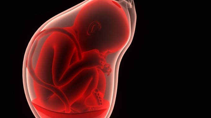
Creating the small intestine, kidney, and lungs of the fetus by culturing cells obtained from the amniotic membrane at 16 to 34 weeks of pregnancy.
‘Fetal stem cells’, which have been subject to ethical debate, appear to be an alternative… Accelerating the identification of causes of congenital malformations
A research team at University College London, UK, succeeded in creating artificial fetal tissue using cells collected from the amniotic sac. He explained that through this, the cause of congenital malformations that occur in the second half of pregnancy can be identified. /Photo = Getty Image Bank They succeeded in artificially creating various organs such as the stomach, intestines, and lungs of a fetus using only cells collected from the amniotic membrane. This is the first time that organ tissue from a fetus in the second half of pregnancy, over 22 weeks old, has been created. It is expected to help reveal the causes of congenital deformities, which have been difficult to research due to ethical issues.
According to the National Health Insurance Corporation, last year’s birth rate in Korea hit an all-time low of 0.72, while the number of babies born with congenital deformities increased. A congenital deformity is a structural abnormality in the body from birth. Research is continuing to determine the cause of the deformity, but it has been difficult to pinpoint because of various genetic and environmental factors. There were also limitations in using organs obtained from actual fetuses.
On the 5th, the research team led by Paolo de Coppi, professor of pediatric surgery at University College London (UCL) in the UK, announced in the international academic journal ‘Nature Medicine’ that they had succeeded in creating artificial fetal tissue using cells collected from the amniotic membrane. Using this, the cause of congenital malformations that occur in the second half of pregnancy can be identified. Additionally, the use of fetal stem cells obtained through abortion can replace existing stem cell technology, which has caused ethical controversy.
The technology to create a human fetal model by culturing stem cells obtained from fetal tissue is fraught with ethical and legal issues. Depending on the country, abortion itself may be illegal, and there is no consensus on whether it is ethically appropriate to use fetal stem cells obtained through abortion for research. Even if stem cells obtained from the fetus are used, only fetal stem cells before the 22nd week of pregnancy can be obtained, so research on fetal development in late pregnancy has been limited.
Professor Kofi’s research team created artificial fetal tissue by culturing epithelial cells obtained from the amniotic membrane of pregnant women at 16 to 34 weeks of age. The amniotic sac is a thin membrane that surrounds the fetus and is filled with amniotic fluid. The research team analyzed the base sequence of the epithelial cells covering the inner surface of the amniotic membrane. Through this, epithelial cells that would grow into the fetus’s stomach, intestines, kidneys, and lungs were identified and cultured separately.
The cultured cells proliferated over two weeks and gradually formed a single tissue. The research team confirmed that the cells form the small intestine, kidney, and lung of the fetus, and that each organ has functional characteristics. This technology was then applied to create an artificial lung for a fetus with congenital diaphragmatic hernia. Congenital diaphragmatic hernia is a congenital disease in which an abnormality occurs in the diaphragm area and part of the stomach moves up into the chest above the diaphragm. As a result, it was confirmed that the symptoms of congenital diaphragmatic hernia appeared in the artificial lung.
An artificial organ (organoid) that functions similar to fetal tissue was created using cells collected from the amniotic membrane rather than an actual fetus. Using artificial organs, we can experiment with various treatments without taking the risk of touching the human body directly. Through this, there is the possibility of developing customized treatments tailored to diseases or physical characteristics.
The research team said, “We have now been able to create fetal organs without fetal stem cells obtained through abortion,” and added, “It will help resolve ethical issues and conduct research on fetal development in late pregnancy that has not been done so far.” The significance of the study was revealed.
A scene from a paper briefing conducted online by the research team, including Professor Paolo de Coppi and Dr. Mattia Gerli, on February 26th. /Photo=Nature Medicine
Related articles to this article
[저작권자 @머니투데이, 무단전재 및 재배포 금지]
2024-03-04 16:00:00
#Artificial #fetus #cells #Changing #landscape #congenital #malformation #research

