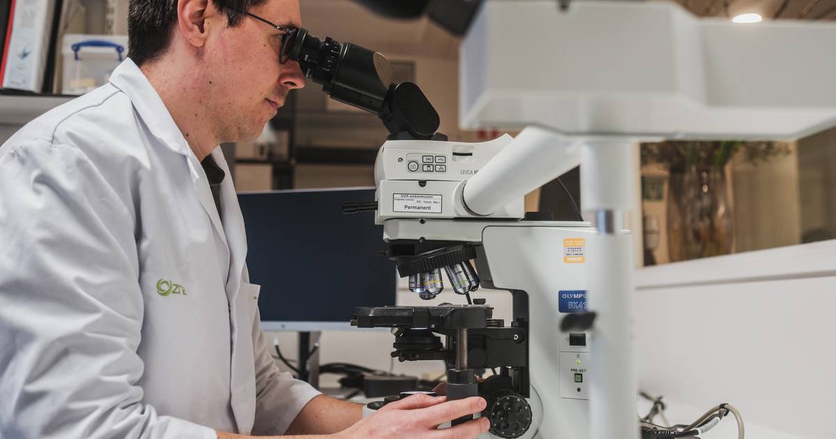
Alzheimer’s disease is the most common form of dementia. It is estimated that one in five Belgians will develop the disease at some point. Researchers have known for some time that the proteins that clump together in the brains of Alzheimer’s patients can form different ‘stems’. These strains determine when the disease starts, how quickly it progresses, and how severe the symptoms are.
Until then, however, the proteins, called amyloid aggregates, have to be removed from the tissues in order to be studied under a particular electron microscope. This makes it difficult to determine how the proteins interact with the rest of the cell and how exactly the different strains arise.
The new microscope solves that problem. “The microscope is linked to infrared spectroscopy and a fluorescence microscope, which allows us to follow the evolution of neuronal degeneration,” says Professor Frederic Rousseau of the VIB-KU Leuven Center for Brain and Disease Research. “Thanks to this new technology, specially designed for our laboratory, we will be able to study the nanoscale structure of strains without removing the amyloid aggregates from the surrounding tissue. This will allow us to study brains that develop symptoms similar to Alzheimer’s from a very young age to follow how different tribes arise.”
In total, the Alzheimer Research Foundation has given more than 4 million euros in financial support to studies on the disease.
SEE ALSO: Can you prevent dementia? Expert explains


