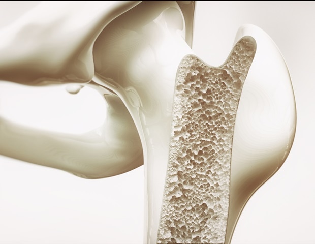
Spatially Resolved Transcriptomic Map Reveals Bone-Muscle Cell Communication
Researchers have developed a spatially resolved transcriptomic map that identifies the precise cellular communication pathways between bone and muscle cells, according to a study detailed by News-Medical. This mapping technique allows scientists to visualize where specific genes are expressed within the physical architecture of the musculoskeletal system, providing a blueprint for how these two tissues coordinate growth and repair.
- Spatial Mapping: The technology captures gene expression while preserving the physical location of cells, unlike traditional sequencing that blends tissues together.
- Intercellular Signaling: The map reveals specific ligands and receptors used by bone and muscle cells to “talk” to one another.
- Clinical Application: This data provides a foundation for treating musculoskeletal atrophy and regenerative medicine.
The musculoskeletal system does not function as a collection of independent parts but as an integrated organ. For decades, the medical community understood that bone and muscle shared a symbiotic relationship, yet the exact molecular “handshakes” occurring at the interface—the periosteum and the endomysium—remained opaque. This gap in knowledge has historically limited the efficacy of treatments for sarcopenia and osteoporosis, where the breakdown of one tissue often accelerates the decline of the other.
The research utilized spatially resolved transcriptomics (SRT), a method that bridges the gap between traditional histology and single-cell RNA sequencing. While standard sequencing tells researchers which genes are active, SRT tells them where those genes are active. This is critical because the pathogenesis of many musculoskeletal disorders is localized to specific niches within the tissue. By mapping these interactions, researchers can identify the exact proteins that trigger muscle regeneration or bone resorption.
How does spatial transcriptomics change the understanding of bone-muscle crosstalk?
Traditional “bulk” sequencing requires grinding tissue into a slurry, which destroys the spatial context and averages the signal across different cell types. According to the findings reported by News-Medical, the new map preserves the anatomical coordinates of the cells. This allows for the identification of “neighborhoods” of cells that work together. For example, the map highlights how osteoblasts in the bone may release signaling molecules that directly influence the metabolic rate of adjacent myofibers in the muscle.


This level of granularity is essential for understanding the standard of care for complex fractures and degenerative joint diseases. When a patient suffers from severe muscle wasting, the morbidity is often compounded by a loss of bone density. Understanding the molecular triggers for this synchronized decay allows for the development of targeted biologics. For patients struggling with these systemic declines, it is highly recommended to consult with [Relevant Clinic/Professional/Service] to explore advanced diagnostic imaging and metabolic screening.
The study’s methodology aligns with broader trends in precision medicine, moving away from systemic drug delivery toward site-specific interventions. By identifying the specific receptors involved in this crosstalk, pharmacological agents can be designed to target only the interface between bone and muscle, potentially reducing systemic side effects and contraindications associated with broad-spectrum steroids or hormones.
Who funded this research and what is the primary source?
The development of these transcriptomic maps is typically the result of multi-institutional collaborations funded by government health agencies and academic grants. While the News-Medical report summarizes the breakthrough, the foundational data is derived from peer-reviewed research focusing on spatial genomics. Much of this work is supported by grants from the National Institutes of Health (NIH) and similar global bodies dedicated to genomic medicine. The integration of this data into clinical practice is currently being vetted through various university-led research portals and the PubMed database to ensure reproducibility across different patient demographics.

The transition from a laboratory map to a clinical tool requires rigorous validation. Current research is moving toward applying these maps to human pathology, specifically examining how the bone-muscle interface changes during aging or after a stroke. This progression follows the trajectory of other “omics” technologies, which first map a healthy state before identifying the biomarkers of disease.
What are the implications for regenerative medicine and B2B healthcare?
The ability to pinpoint cellular communication opens a new door for tissue engineering. If scientists can replicate the signaling environment identified in the transcriptomic map, they can more effectively grow synthetic bone and muscle grafts that integrate seamlessly with a patient’s existing anatomy. This reduces the risk of graft rejection and improves the functional outcome of reconstructive surgeries.

From a business and regulatory perspective, this shift toward spatial biology requires a new infrastructure for diagnostics. Pathology labs must now integrate high-resolution imaging with genomic sequencing. This technological leap means that diagnostic centers are actively seeking [Relevant Clinic/Professional/Service] to implement these complex workflows while remaining compliant with evolving healthcare data regulations. Furthermore, pharmaceutical companies developing the next generation of musculoskeletal drugs are prioritizing “spatial biomarkers” to prove efficacy in clinical trials.
The clinical logic is clear: if you can see the conversation between cells, you can interrupt a pathological signal or amplify a healing one. This is particularly relevant for the treatment of chronic conditions where the standard of care has plateaued. By leveraging these maps, providers can move toward a “personalized musculoskeletal profile” for each patient, tailoring the treatment to the specific molecular deficiencies of their bone-muscle interface.
As this research moves toward broader clinical application, the focus will shift toward identifying the specific ligands that can be synthesized into therapeutic drugs. The eventual goal is a pharmacological “switch” that can trigger the bone-muscle communication network to accelerate healing after trauma. To stay ahead of these developments, healthcare providers and patients should utilize vetted directories to connect with [Relevant Clinic/Professional/Service] specializing in regenerative orthopedics and genomic medicine.
Disclaimer: The information provided in this article is for educational and scientific communication purposes only and does not constitute medical advice. Always consult with a qualified healthcare provider regarding any medical condition, diagnosis, or treatment plan.