Fishbone Ingestion Causing Cecal Perforation: A Case Report
A patient underwent a diagnostic laparoscopic appendectomy that instead revealed a cecal perforation caused by an ingested fishbone, according to a case report. The finding demonstrates how sharp dietary foreign bodies can lodge in the gastrointestinal tract and produce symptoms that closely resemble acute appendicitis.
- A patient presenting with acute right lower quadrant abdominal pain was found to have a fishbone perforation in the cecum rather than standard acute appendicitis.
- Preoperative contrast-enhanced computed tomography failed to definitively visualize the small fishbone prior to surgical exploration.
- Laparoscopy allowed for successful foreign body retrieval, oversewing of the cecal perforation, and appendectomy without postoperative complications.
Emergency Room Presentation and Initial Evaluation
The patient arrived at the emergency room reporting right lower quadrant abdominal pain. The pain started suddenly at a moderate intensity and escalated over time, accompanied by a loss of appetite and nausea. Physical examination revealed moderate tenderness in the right lower abdomen alongside rebound tenderness and mild rigidity. Vital signs remained within normal ranges.
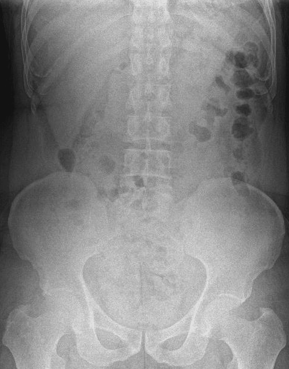
Laboratory evaluations showed a hemoglobin level of 14.2 grams per deciliter, a white blood cell count of 11,000 per cubic millimeter with 64.6 percent neutrophils, a platelet count of 230,000 per cubic millimeter, a blood urea nitrogen level of 14.5 milligrams per deciliter, and a serum creatinine level of 0.68 milligrams per deciliter. A plain abdominal radiograph showed no radiopaque foreign body.
CT Scan Suggests Appendicitis but Misses Foreign Body
Because plain X-rays lack sensitivity for low-radiopacity foreign bodies, clinicians ordered a contrast-enhanced computed tomography scan of the abdomen. The imaging revealed a non-aerated appendix measuring six millimeters in diameter with enhancing walls and multiple surrounding reactive lymph nodes. Mild surrounding fat stranding, minimal free fluid, and mild mural thickening of the cecum were also noted.
Although the imaging pointed toward potential acute appendicitis, the scan did not definitively identify a foreign body. Medical literature notes that small fishbones can be missed on initial radiological review due to slice thickness, bowel peristalsis, and the orientation of the object relative to axial slices. Consequently, surgical exploration was scheduled.
Laparoscopic Discovery and Surgical Intervention
Under general anesthesia, the surgical team performed a diagnostic laparoscopy using an open technique for pneumoperitoneum. Exploration revealed a grade I inflamed appendix that was hyperemic and swollen in its distal half. Upon mobilizing the appendix for removal, the surgeon identified a sharp white foreign body penetrating the anterior wall of the cecum and adhering to the appendix.
The surgical team cut the appendix between loop ligatures and removed it. Clinicians retrieved the foreign body using forceps and closed the punctate perforation with a figure-of-eight Vicryl 3-0 suture. Histopathological examination of the removed appendix confirmed intact mucosa and multiple reactive lymphoid follicles, indicating secondary inflammation driven by the neighboring cecal perforation rather than primary appendicular pathology.
The patient recovered without incident, tolerated an oral diet after drain removal on the second postoperative day, and was discharged on the third postoperative day.
Disclaimer: The information provided in this article is for educational and scientific communication purposes only and does not constitute medical advice. Always consult with a qualified healthcare provider regarding any medical condition, diagnosis, or treatment plan.