Intraoperative Right Coronary Artery Occlusion During Hip Hemiarthroplasty: A Case Report
An 86-year-old woman undergoing right hip hemiarthroplasty suffered a silent intraoperative right coronary artery occlusion at a prior stent site, progressing to ventricular fibrillation during emergency transfer. The case details a timeline where continuous electrocardiographic monitoring captured progressive ST-segment elevation before any overt hemodynamic collapse, prompting rapid nursing verification, multidisciplinary intervention, and successful percutaneous coronary intervention.
Key Clinical Takeaways:
- An 86-year-old female patient developed an acute intraoperative right coronary artery occlusion at a prior stent site during hip hemiarthroplasty under spinal anesthesia.
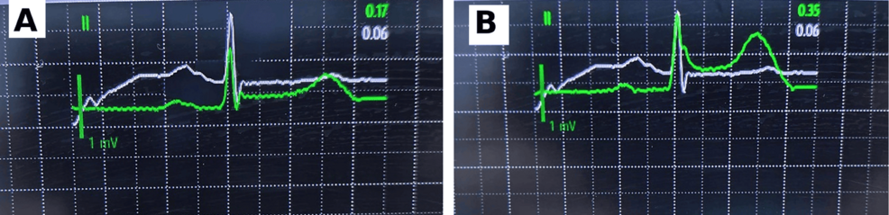
- Continuous ECG monitoring detected progressive ST-segment elevation in lead II from 0.17 mV to 0.35 mV, recognized and verified by the anesthesia nurse before hemodynamic deterioration.
- Emergency coronary angiography and drug-eluting stent placement restored TIMI grade 3 flow after an episode of ventricular fibrillation during transfer, leading to patient survival and discharge on postoperative day 15.
Intraoperative Ischemia During Hip Hemiarthroplasty
Hip fracture surgery in older adults carries substantial perioperative cardiovascular risk, with a reported 30-day myocardial infarction prevalence of approximately 2% according to recent systematic reviews. Patients with previous coronary stent implantation who require urgent non-cardiac surgery represent a complex clinical challenge, as antiplatelet therapy is often interrupted to balance bleeding and thrombotic risks. In this case, the patient had discontinued clopidogrel one week prior to admission for her right femoral neck fracture. Her medical history included hypertension and coronary artery disease, having undergone percutaneous coronary intervention with a right coronary artery stent. Preoperative electrocardiography showed sinus rhythm with ST-T abnormalities in inferior leads.
Nurse Alerts Team After Monitor Shows ST-Segment Elevation
Surgery began at 09:00 hours following the administration of spinal anesthesia with 2.0 mL of 0.5% hyperbaric ropivacaine, which achieved a sensory block fixed at T10 by 08:20. At 09:32, the perioperative monitor registered progressive ST-segment elevation. Review of the stored ST-analysis data revealed a rise in lead II from roughly 0.17 mV at 09:33:06 to 0.35 mV by 09:37:06. The anesthesia nurse recognized the abnormal trend, verified the monitor findings, and immediately alerted the anesthesiology team while initiating an urgent cardiology consultation at approximately 09:33. Heart rate and blood pressure began to fall at 09:34, prompting the administration of intravenous nitroglycerin, norepinephrine, and dopamine, alongside invasive arterial pressure monitoring.
Patient Suffers Ventricular Fibrillation During Transfer for Angiography
A 12-lead ECG obtained between 10:10 and 10:15 was interpreted clinically as an acute inferior myocardial infarction, leading to the decision to proceed with emergency transfer for coronary angiography at approximately 10:40. Between 10:41 and 10:45, the patient experienced ventricular fibrillation during transit to the catheterization laboratory. Nursing staff performed cardiopulmonary resuscitation and delivered electrical defibrillation at 200 J per attempt. While initial shocks produced only transient rhythm recovery with recurrent ventricular fibrillation, an organized rhythm was documented at approximately 10:56. Coronary angiography revealed an acute proximal-to-mid right coronary artery occlusion at the previous stent site accompanied by thrombus. Following the delivery of intracoronary tirofiban and the deployment of a 2.75 × 29 mm drug-eluting stent, balloon angioplasty reestablished TIMI grade 3 flow, leaving about 30% residual stenosis.
Cardiac Biomarkers Decline Before Patient Is Discharged
Postoperative cardiac biomarker evaluation confirmed significant myocardial injury followed by gradual decline. On postoperative day 1, troponin T exceeded 50 ng/mL, CK-MB measured 130.51 ng/mL, myoglobin reached 400.64 mg/mL, and BNP registered 8,994.37 ng/mL. By postoperative day 5, troponin T rose to 4,056 ng/mL before declining to 900 ng/mL by day 9. The patient experienced no major bleeding, transfusions, or neurological complications, and successfully ambulated prior to discharge on postoperative day 15. The clinical report emphasizes that while continuous surveillance provided objective evidence of evolving ischemia, the observational single-case chronology does not establish that any specific monitoring or escalation strategy alters clinical outcomes.