How Vitamin A and Thyroid Hormones Shape Human Vision: A Breakthrough Discovery
Scientists at Johns Hopkins University published in the Proceedings of the National Academy of Sciences that a vitamin A derivative works alongside thyroid hormones to form the foveola, the tiny center of the human retina responsible for sharp central vision. The research challenges a thirty-year-old scientific model of how human color vision develops before birth.
The Tech TL;DR:
- Researchers found that retinoic acid (from vitamin A) and thyroid hormones cause early blue-light-detecting cells in the fetal foveola to transform into red- and green-light-detecting cones.
- The discovery overturns the three-decade-old assumption that initial cone cells simply migrate away or remain permanently fixed in their early identity.
- Laboratory-grown retinal organoids from fetal cells enabled the team to track human-specific retinal development that traditional animal models like mice or fish cannot replicate.
Fetal Cell Transformation Discovered in Lab-Grown Retinal Organoids
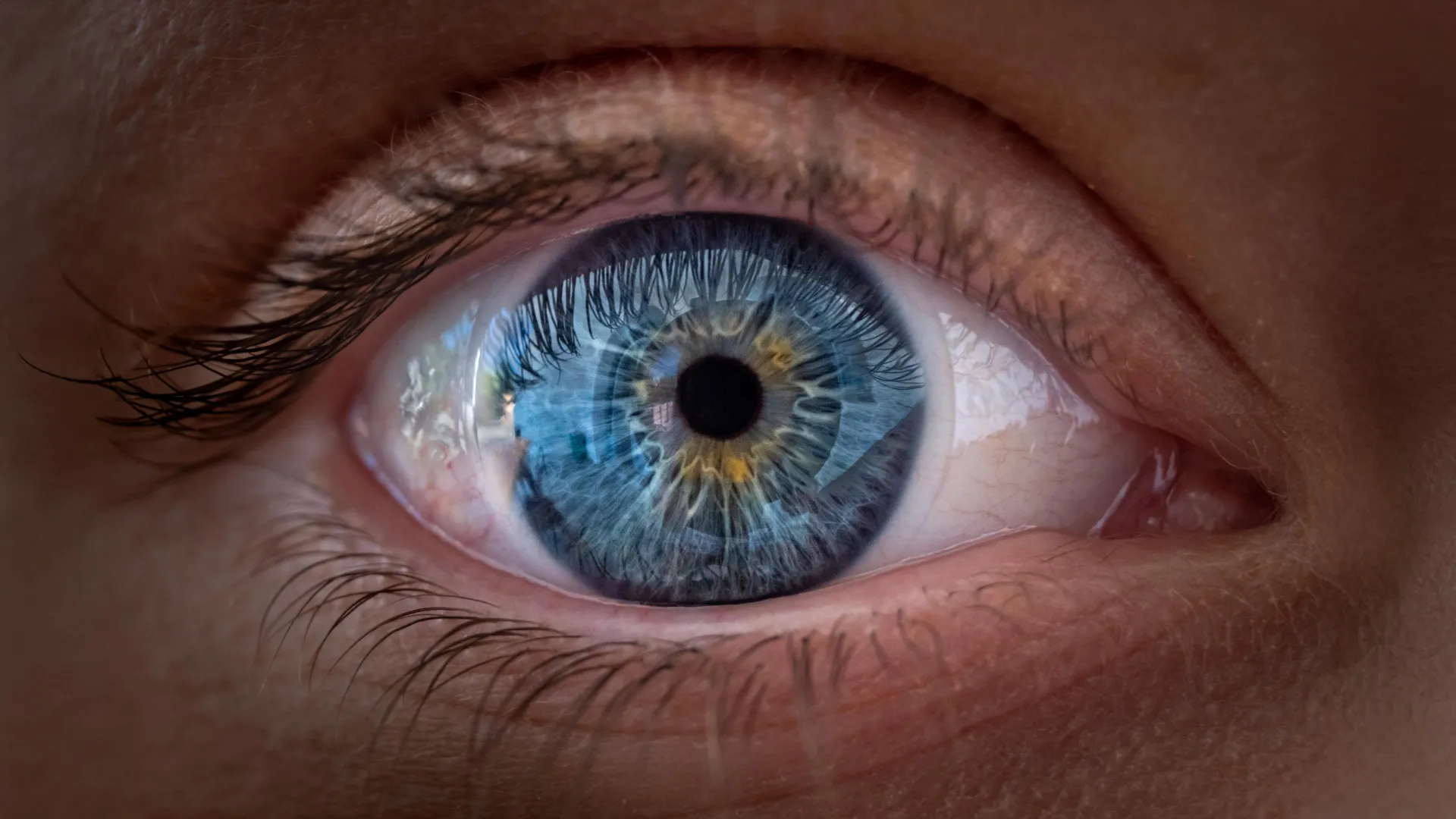
Investigating human vision formation required an innovative laboratory technique because commonly studied animals such as mice and fish do not share the specific retinal organization found in humans. Robert J. Johnston Jr., an associate professor of biology at Johns Hopkins University who led the study, and his colleagues grew miniature versions of human retinal tissue called organoids from fetal cells to observe development over several months. Their attention centered on the foveola, a tiny region at the very center of the retina that occupies only a small fraction of the tissue but is involved in approximately 50 percent of human visual perception. Within the foveola, specialized light-detecting cells called cones support daytime vision and color perception. While typical retinal regions contain blue, green, and red cones, the foveola contains red and green cones but lacks blue cones. This precise arrangement creates the sharp central vision required to read, recognize faces, and distinguish fine details.
Vitamin A Breakdown and Thyroid Hormones Shape Central Vision
The research team tracked two coordinated processes during weeks 10 through 12 of fetal development when a small number of blue cones are initially present in the foveola. By week 14, those cells transform into red and green cones. Two chemical influences drive this conversion. First, retinoic acid, a molecule produced from vitamin A, breaks down to limit the formation of blue cones in the central region. Second, thyroid hormones encourage the remaining blue cones to change into red and green cones. First, retinoic acid helps set the pattern. Then, thyroid hormone plays a role in converting the leftover cells,
Johnston stated in the findings. That’s very important because if you have those blue cones in there, you don’t see as well.
The discovery concerns how vitamin A signaling guides eye development before birth, rather than establishing that vitamin A supplements improve sharp vision in adults.
Challenging a Thirty-Year-Old Scientific Theory of Cone Migration
The results offer an alternative to an explanation that has influenced vision research for decades. Under the previous model, scientists assumed that the few blue cones initially appearing in the foveola simply moved away as the retina developed, and that once cells became a specific type of cone, their identities remained fixed forever. The main model in the field from about 30 years ago was that somehow the few blue cones you get in that region just move out of the way, that these cells decide what they’re going to be, and they remain this type of cell forever,
Johnston noted. We can’t really rule that out yet, but our data supports a different model. These cells actually convert over time, which is really surprising.
While cell migration cannot be entirely ruled out, the observations indicate that developing cone cells possess greater flexibility than previously recognized.
Implications for Treating Macular Degeneration and Restoring Lost Vision
Beyond explaining healthy eye development, the findings point toward future treatments for macular degeneration, a major cause of central vision loss among older adults that damages the macula without a current cure. Johnston’s team is working to refine retinal organoids to better replicate human eye structure and function, with the long-term goal of producing precisely controlled populations of photoreceptors for transplantation into damaged retinas. Katarzyna Hussey, a study author, former doctoral student in Johnston’s laboratory, and current molecular and cell biologist at CiRC Biosciences in Chicago, outlined the therapeutic trajectory. The goal with using this organoid tech is to eventually make an almost made-to-order population of photoreceptors,
Hussey said. A big avenue of potential is cell replacement therapy to introduce healthy cells that can reintegrate into the eye and potentially restore that lost vision.
Hussey emphasized that clinical application remains distant, requiring extensive safety and efficacy optimizations before human trials can begin.