Eyebrow follicular mucinosis reveals classic Hodgkin lymphoma
Eyebrow Lesion Reveals Classic Hodgkin Lymphoma
A 26-year-old man who developed a scaly plaque on his right eyebrow in December 2024 ultimately received a diagnosis of classic Hodgkin lymphoma, nodular sclerosis subtype, after doctors investigated the unusual skin finding alongside persistent cervical lymphadenopathy and weight loss.
-
Key Clinical Takeaways:
- Follicular mucinosis of the eyebrow can act as an early cutaneous marker for an underlying systemic lymphoproliferative disorder.
- Dermoscopy revealed a characteristic “toothpaste sign” with whitish material protruding from follicular ostia, guiding the initial skin biopsy in January 2025.
- Systemic chemotherapy with doxorubicin, bleomycin, vinblastine, and dacarbazine led to a complete metabolic response and full eyebrow hair regrowth by late 2025.
Patient Develops Scaling and Hair Loss on Eyebrow
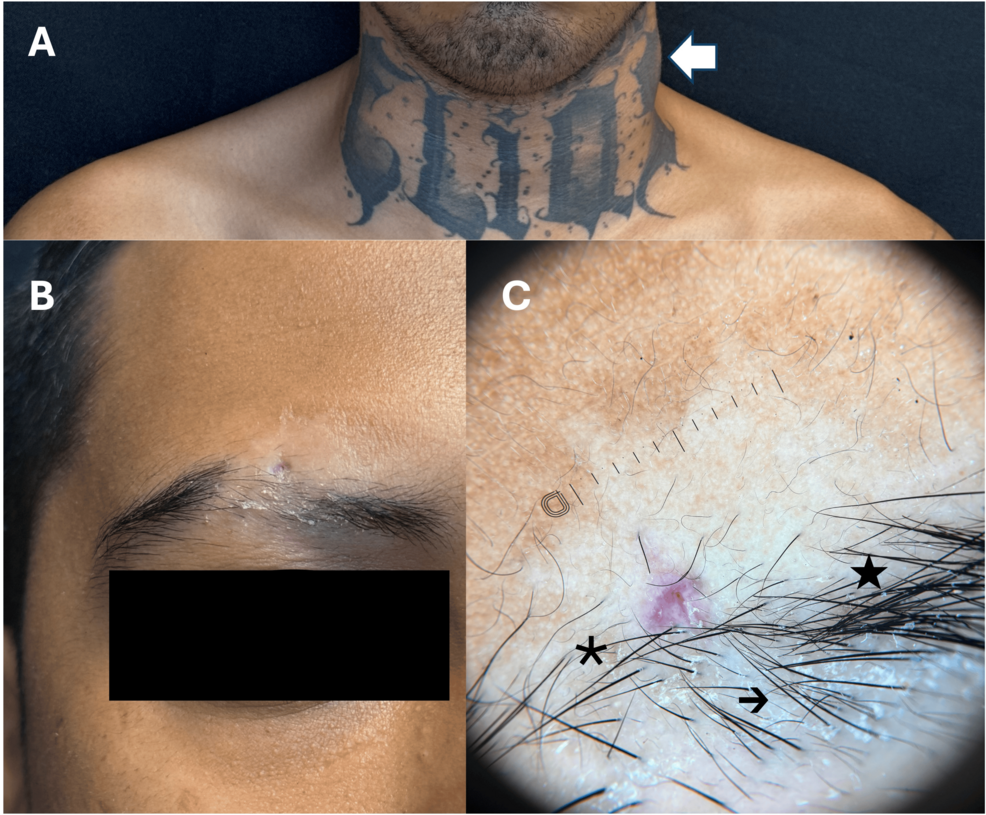
The patient initially noticed localized scaling on his right eyebrow in December 2024, followed one week later by progressive hair loss in the affected region. Approximately one month after the initial symptom, a hypopigmented patch emerged. When clinicians examined the area, they observed a 2.2 by 2.0 centimeter hypopigmented, infiltrated, and pseudoalopecic plaque possessing irregular borders across the central third of the right eyebrow. Cervical lymphadenopathy was also documented during the initial physical evaluation. Dermoscopy showed reduced hair density, hairs oriented in multiple directions, central whitish scale, and distinct whitish material protruding through several follicular ostia, a clinical indicator designated as the toothpaste sign. Initial differential diagnoses considered by the medical team included alopecia areata, lichen sclerosus, and seborrheic dermatitis. Because the plaque was infiltrated and accompanied by follicular scaling, a skin biopsy was performed in late January 2025.
Biopsy Confirms Follicular Mucinosis with Mucin Deposition
Histopathologic analysis of the eyebrow lesion demonstrated reticular degeneration within the follicular epithelium, marked by the separation of outer root sheath keratinocytes due to pale mucinous material. A sparse perifollicular and perivascular lymphocytic inflammatory infiltrate appeared in the dermis. Staining with Alcian blue confirmed follicular and interstitial mucin deposition, establishing the definitive diagnosis of follicular mucinosis. Laboratory evaluations revealed a mild anemia with a hemoglobin level of 12.2 grams per deciliter, an elevated lactate dehydrogenase level of 268.7 units per liter against a reference range of 135 to 225 units per liter, and an albumin level of 4.0 grams per deciliter. Complete blood counts, serum chemistry, and liver function tests showed no other abnormalities.
Systemic Staging and Lymph Node Biopsy
Because the patient presented with cervical lymphadenopathy and unquantified weight loss, physicians ordered a contrast-enhanced computed tomography scan of the neck, chest, and abdomen. The imaging uncovered cervical, anterior mediastinal, subcarinal, and lower paratracheal lymphadenopathy, which included a right cervical nodal conglomerate measuring 29 by 37 millimetres and a prevascular mediastinal conglomerate measuring 63 by 14 millimetres. These mediastinal masses caused approximately 50 percent narrowing of the superior vena cava. An excisional lymph node biopsy performed in late April 2025 revealed an atypical lymphoid proliferation featuring a nodular architecture divided by broad fibrous bands. Large neoplastic cells containing binucleated structures and prominent nucleoli appeared within a mixed inflammatory background, matching the morphological profile of Reed-Sternberg cells. Immunohistochemical staining demonstrated membranous positivity for CD30 and CD15, weak granular expression of CD20, and negativity for CD3 and Epstein-Barr virus latent membrane protein 1. These results confirmed a diagnosis of classic Hodgkin lymphoma, nodular sclerosis subtype, categorized as Ann Arbor stage IIB.
Chemotherapy Resolves Eyebrow Symptoms and Residual Nodes Remain
Following the hematologic staging, the patient began combination chemotherapy in late April 2025 utilizing the doxorubicin, bleomycin, vinblastine, and dacarbazine regimen. By the fifth cycle administered in October 2025, the hypopigmentation, tissue infiltration, and scaling of the eyebrow had completely resolved, accompanied by full regrowth of eyebrow hair. The patient finished a total of seven chemotherapy cycles in December 2025. Subsequent positron emission tomography and computed tomography scans showed residual mediastinal lymph nodes measuring up to 27 millimetres with a maximum standardized uptake value of 1.7, corresponding to a complete metabolic response with a Lugano-Deauville score of 2. Follow-up evaluations continued through July 2026 under the care of the hematology department.
Disclaimer: The information provided in this article is for educational and scientific communication purposes only and does not constitute medical advice. Always consult with a qualified healthcare provider regarding any medical condition, diagnosis, or treatment plan.