DLBCL Diagnosed in 19-Year-Old Male, Cureus Reported
A 19-year-old male patient presented with a three-month history of a painless, progressively enlarging mass in his left groin, leading to a diagnosis of stage IA diffuse large B-cell lymphoma (DLBCL), Cureus reported.
- A 19-year-old male developed an isolated 6 cm left inguinal mass over three months without experiencing B symptoms like fever or night sweats.
- The patient achieved complete metabolic remission following three cycles of R-CHOP chemotherapy and proton beam therapy.
Clinical Presentation of Inguinal Diffuse Large B-Cell Lymphoma
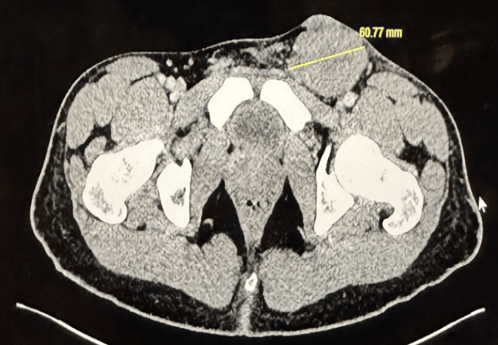
Diffuse large B-cell lymphoma typically affects older adults and rarely presents in individuals under the age of 20, accounting for approximately 1.3% of new cases in Surveillance, Epidemiology, and End Results data cited by Cureus. The 19-year-old patient sought care for a firm, non-tender, mobile 5 by 6 cm swelling in the left inguinal region. He had received a course of oral antibiotics from a primary care provider with no improvement, and he reported no systemic B symptoms such as unexplained weight loss or night sweats.
Imaging and Biopsy Reveal Atypical Lymphoid Cells
Computed tomography demonstrated a 6.4 by 5.1 cm heterogeneous superficial soft tissue lesion in the left groin extending to the skin surface, accompanied by low-volume reactive lymph nodes and fat stranding. Radiologists initially included inflamed sebaceous cysts, hematomas, and Kimura disease in the differential diagnosis. A core needle biopsy of the left lymph node subsequently revealed architecture effaced by atypical lymphoid cells displaying a B-cell phenotype positive for CD20, CD10, BCL6, and BCL2, alongside a high proliferative index of approximately 70% on MIB-1 staining. RNA sequencing confirmed a BCL2::IGH fusion, classifying the disease as germinal center type DLBCL.
PET Scan Confirms Stage IA Disease
Baseline fluorodeoxyglucose positron emission tomography-computed tomography identified a 5.7 cm FDG-avid left inguinal mass with a maximum standardized uptake value of 21.8. Additional PET-avid skin lesions on the chest wall and axilla showed no lymphoma involvement upon biopsy, while bone marrow and cerebrospinal fluid evaluations remained negative. Clinicians documented an elevated lactate dehydrogenase level, contributing to an International Prognostic Index score of 1 and staging the disease as stage IA. Following negative viral screenings and sperm cryopreservation, the patient received three cycles of rituximab, cyclophosphamide, doxorubicin, vincristine, and prednisone, known as R-CHOP, followed by proton beam therapy to the left inguinal region.
Patient Achieves Complete Metabolic Remission
End-of-treatment positron emission tomography confirmed complete metabolic remission after the combined immunochemotherapy and proton beam therapy regimen. The patient returned to his baseline functional status while managing transient, localized radiotherapy side effects including reduced hair growth and mild discomfort. Hematology teams established a five-year follow-up schedule in the lymphoma clinic to monitor for potential disease recurrence and long-term treatment-related complications.
Disclaimer: The information provided in this article is for educational and scientific communication purposes only and does not constitute medical advice. Always consult with a qualified healthcare provider regarding any medical condition, diagnosis, or treatment plan.