Scalp tumor from burn scar invades brain, Cureus reports
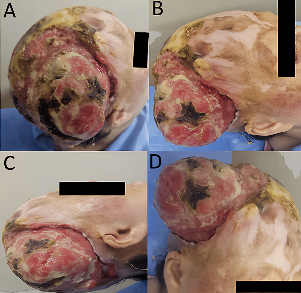
A 38-year-old female patient presented to clinicians with a large, exophytic scalp tumor that had silently invaded her skull and brain tissue without triggering a single neurological deficit, Cureus reported. The malignant lesion arose from a thermal burn scar sustained 33 years prior, highlighting an extreme case of a chronic wound transformation where extensive intracranial destruction developed while cognitive and motor functions remained entirely intact.
Key Clinical Takeaways:
- An ulcer developed in a childhood thermal burn scar after a 33-year latency period, eventually eroding through the skull into the dura mater and cerebral parenchyma.
- Despite severe intracranial invasion, the patient maintained a Glasgow Coma Scale score of 15/15 and showed no focal neurological deficits at initial presentation.
- Complete surgical resection was deemed unfeasible due to the massive tissue defect required, and the patient ultimately declined definitive chemoradiotherapy.
Thirty-Three Years of Latency Followed by Accelerated Growth
The patient sustained a second-degree thermal burn at three years of age, which healed slowly and left an area of scarring alopecia and pigment changes. Approximately 33 years later, an ulcer emerged within the old scar tissue. While the lesion grew slowly at first, its expansion rate accelerated sharply during the final three months before clinical evaluation. A persistent foul odor ultimately prompted the patient to seek medical care.
Physical examination revealed a multilobulated, ulcerated, and exophytic tumor covering a vast portion of the right scalp. The mass was friable, fixed to underlying structures, and coated with fibrinous exudate alongside regions of necrosis. Blood tests demonstrated neutrophilic leukocytosis alongside a C-reactive protein level of 130 mg/L. A wound swab identified a Staphylococcus aureus infection sensitive to all tested antibiotics, which clinicians managed successfully with a 10-day course of amoxicillin-clavulanate and local wound care.
Imaging Reveals Massive Scalp Mass and Skull Destruction
Initial brain computed tomography imaging exposed a massive right-sided scalp mass accompanied by profound destruction of the skull bone. The scans captured thickening of the underlying dura mater and outward herniation of brain tissue through the osseous defect. A subsequent dural biopsy yielded a histological diagnosis of well-differentiated, infiltrating, keratinizing squamous cell carcinoma characterized by keratin pearls and tumor necrosis.
Magnetic resonance imaging performed two weeks later mapped the full extent of the pathology, demonstrating a right temporoparieto-occipital scalp tumor stretching 11.5 centimeters with a maximum thickness of 12 millimeters. The imaging confirmed direct infiltration into the adjacent right temporoparietal cerebral parenchyma, accompanied by a 55 by 35 millimeter intraparenchymal hematoma and marked perilesional edema. Staging via cervico-thoraco-abdominopelvic computed tomography revealed no regional lymph-node involvement or distant metastases, placing the disease at cT4aN0M0 under American Joint Committee on Cancer criteria.
Patient Declines Chemoradiotherapy After Surgery Is Deemed Impractical
The multidisciplinary tumor board determined that surgical resection was impractical given the profound skin, calvarial, dural, and cerebral involvement. Clinicians proposed a definitive concurrent chemoradiotherapy regimen consisting of 66 grays delivered in 33 fractions alongside weekly cisplatin. However, the patient declined this treatment path. Two months later, she returned with severe headaches, vomiting indicative of intracranial hypertension, and new facial paralysis.
A noncontrast brain computed tomography scan showed stable tumor dimensions paired with worsening perilesional edema and mass effect. Following intravenous corticosteroid boluses, she completed a palliative course of three-dimensional conformal radiotherapy totaling 30 grays across 10 fractions. Although early tolerance was acceptable, her symptoms failed to improve, and she died approximately one month after completing palliative irradiation.
Disclaimer: The information provided in this article is for educational and scientific communication purposes only and does not constitute medical advice. Always consult with a qualified healthcare provider regarding any medical condition, diagnosis, or treatment plan.