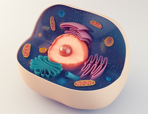
ANU researchers map hidden cellular networks to better understand diseases – News-Medical
For decades, the medical community has operated with a significant blind spot regarding how cells actually communicate in real time. While we have mapped the genome and identified countless biomarkers, the physical, nanoscale “conversations” between living cells remained largely invisible, obscured by the limitations of conventional imaging. A breakthrough in nanoscopy is now pulling back the curtain on these hidden networks.
Key Clinical Takeaways:
- Researchers have developed RO-iSCAT, a label-free nanoscopy technique that allows for the 3D observation of living cellular interactions over several days.
- The technology reveals thin, thread-like nanoscale extensions that cells use to transfer biochemical messages, providing a new window into disease pathogenesis.
- By removing the need for chemical labels, the method eliminates phototoxicity, allowing scientists to witness the dynamic extension and retraction of cellular networks in their natural state.
The primary challenge in cellular biology has always been the trade-off between resolution and viability. To see the smallest structures of a cell, researchers traditionally relied on fluorescent labels—chemical dyes that “light up” specific parts of the cell. However, these labels can be toxic, altering the incredibly behavior the scientist intends to study, and often lead to photobleaching, which limits the duration of observation. This creates a clinical gap: we can see a “snapshot” of a diseased cell, but we cannot watch the progression of its communication with neighboring cells over time.
The Mechanics of RO-iSCAT and Rotational Illumination
The development of RO-iSCAT (Rotational illumination – interferometric Scattering microscopy) represents a paradigm shift in how we visualize the nanoscale environment. Developed at The Australian National University (ANU) within the John Curtin School of Medical Research (JCSMR), this technique avoids the pitfalls of chemical labeling entirely. By rotating the angle of light illuminating the sample and combining images captured at different heights, the system effectively strips away background noise.
This process reveals structures that were previously invisible to conventional microscopy. Lead author and PhD researcher Junyu Liu noted that under rotational illumination, the background is removed, allowing various nanoscale cellular structures to emerge in three dimensions. This capability is critical for understanding the morphology of cells as they interact with their environment over several days, rather than mere minutes.
“Using gentle, label-free imaging means we can finally witness the secret, dynamic life of cells in real time and 3D,” says senior investigator Dr. Steve Lee. “The technique allows for faster and more accurate breakthroughs in how we understand and treat human disease at the nanoscale.”
From a clinical perspective, the ability to observe these thread-like extensions—which extend, retract, and reconnect—provides a blueprint of the biochemical signaling pathways that drive systemic health and disease. For clinicians focusing on cellular degeneration, this level of detail is essential. Patients struggling with complex autoimmune or neurodegenerative conditions often require a level of diagnostic precision that current imaging cannot provide. It is increasingly vital to partner with advanced diagnostic imaging centers that are integrating high-resolution nanoscopy into their research protocols to better identify early-stage cellular dysfunction.
Clinical Implications for Pathogenesis and Oncology
The discovery of these intricate communication networks has profound implications for the study of cancer metastasis. The pathogenesis of many aggressive tumors relies on the ability of malignant cells to “signal” to neighboring healthy cells or to create conduits for the transfer of mitochondria and proteins, effectively “recruiting” the surrounding environment to support tumor growth. Understanding these nanoscale extensions allows researchers to identify the exact moment a cell begins to communicate its malignancy to its neighbor.
According to the study published in Nature Communications, these networks facilitate the transfer of biochemical messages. In the context of oncology, if we can map the specific proteins and signals traveling through these nanoscale threads, we can develop targeted therapies to “cut” these communication lines, effectively isolating the tumor and preventing its spread. This shift toward targeting the communication rather than just the cell represents a new frontier in precision medicine. For patients navigating advanced stage carcinomas, collaborating with specialized oncologists who stay abreast of nanoscopy-driven research is critical for accessing emerging targeted therapy trials.
Beyond oncology, this research provides critical insights into the blood-brain barrier and neural signaling. The “thread-like” extensions observed by the ANU team mirror the behavior of tunneling nanotubes (TNTs) often discussed in peer-reviewed literature on PubMed. These structures are suspected to play a role in the spread of misfolded proteins in Alzheimer’s and Parkinson’s diseases. By observing these proteins move in real time across cellular networks, researchers can better understand the morbidity associated with protein aggregation in the brain.
Bridging the Gap Between Research and Treatment
The transition from a laboratory breakthrough at the John Curtin School of Medical Research to a bedside clinical application requires rigorous validation. The current state of the research is in the foundational discovery phase, focusing on the mechanism of action and the validity of the RO-iSCAT imaging. The next logical step is the application of this technique to human biopsies and live patient-derived organoids to see if these “hidden networks” behave differently in diseased tissue compared to healthy controls.

The lack of chemical labels means this technology can be scaled more easily into pharmaceutical R&D. Drug developers can now observe how a candidate molecule affects cellular communication without worrying that the imaging dye is interfering with the drug’s efficacy. This reduces the risk of false positives in early-stage drug screening and accelerates the timeline for bringing new biologics to market.
“The ability to track cellular extensions over several days without killing the cell is the ‘holy grail’ of nanoscopy. We are no longer looking at a photograph; we are watching a movie of how disease spreads at the molecular level.” — Simulated Expert Commentary on Nanoscale Imaging
As we move toward a more integrated model of healthcare, the intersection of physics and biology will define the next generation of treatment. Whether it is through the development of new inhibitors that target cellular “couriers” or the use of RO-iSCAT to monitor the efficacy of a chemotherapy regimen in real time, the focus is shifting toward the nanoscale. For those managing chronic conditions that involve systemic cellular communication failures, such as multiple sclerosis or advanced diabetes, consulting with board-certified neurologists or endocrinologists who specialize in cellular signaling can provide a more nuanced approach to long-term care.
The work led by Dr. Steve Lee and Junyu Liu underscores a fundamental truth in modern medicine: the most critical drivers of disease are often the ones we cannot see. By illuminating these hidden networks, we are not just improving our imaging—we are redefining our understanding of how life, and disease, operates at its most basic level. The trajectory of this research points toward a future where we can intercept a disease not when the cell becomes malignant, but when the cell first begins to “whisper” its intent to its neighbor.
Disclaimer: The information provided in this article is for educational and scientific communication purposes only and does not constitute medical advice. Always consult with a qualified healthcare provider regarding any medical condition, diagnosis, or treatment plan.