
CLEVELAND – Researchers at Case Western Reserve University have achieved a significant breakthrough in cancer treatment by developing a method to overcome one of the most challenging obstacles: the dense physical barrier that solid tumors construct around themselves, hindering the delivery of therapeutic drugs.
The team, led by Efstathios “Stathis” Karathanasis, vice chair and professor of biomedical engineering, and Agata Exner, the Henry Willson Payne Professor of Radiology, successfully used ultrasound-activated nanobubbles to break down these tumor walls, allowing treatment-bearing molecules to reach the cancer cells within. The findings, published in ACS Nano, offer a potential pathway to improve the efficacy of existing and emerging cancer therapies.
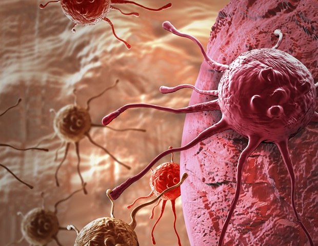
Solid tumors are characterized by a stiff, dense extracellular matrix composed largely of collagen, a protein found in scar tissue. This physical barrier prevents immune cells and modern immunotherapies, particularly RNA carried in lipid nanoparticles, from effectively penetrating the tumor core. “The physical barrier is limiting delivery of cancer drugs, especially for new immunotherapies,” Karathanasis said.
The researchers injected nanobubbles, filled with an inert gas called perfluoropropane, directly into tumors in a breast cancer model. Subsequently, they applied ultrasound waves to gently “jiggle” the bubbles, disrupting the collagen network without causing cellular damage. This process effectively softens the tumor microenvironment, making it more permeable to therapeutic agents and immune cells.
“We drop the defenses of the cancer and give a fair chance for our therapies to actually win,” explained Exner, who also directs the CWRU Center for Imaging Research. “We didn’t invent a new drug, but it has the potential to make any existing or emerging therapy work much better.”
The treatment also demonstrated the ability to activate immune cells already present within the tumors, even without additional therapies. According to Karathanasis, these activated immune cells begin secreting danger signals, attracting more immune cells to the tumor site. Killer T cells targeting the cancer demonstrated the ability to seek out and attack other tumors, even those not directly treated.
The nanobubble treatment maintained the softened state of the tumors for at least five days, while untreated tumors continued to stiffen. When researchers subsequently injected lipid nanoparticles containing RNA to enhance T cell activity, the treatment dispersed throughout the tumor, rather than remaining localized at the injection site.
The potential for rapid clinical translation is high, as the nanobubbles are already being commercialized by Visano Theranostics, a company co-founded by Exner, for prostate cancer detection. Ultrasound, a key component of the therapy, is already FDA-approved and widely available. Exner indicated that an Investigational New Drug (IND) application will be submitted to the FDA within the next 18 months, with the possibility of leveraging that application to expedite clinical trials within two years.
“Any tumor that you can biopsy can potentially have nanobubbles introduced,” Exner said. “This is especially important for solid tumors that are difficult to treat, where ultrasound is already used, like liver, prostate and ovarian cancers.”
The research was supported by funding from the Case Comprehensive Cancer Center and the National Institutes of Health.