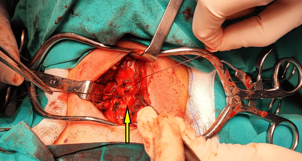
Woman has tracheal laceration and endotracheal tube cuff rupture during thyroidectomy
A 59-year-old woman undergoing an elective right hemithyroidectomy experienced a rare concurrent anterior tracheal laceration and electromyographic endotracheal tube cuff rupture approximately 100 minutes after anesthesia induction. The surgical intervention involved a 7.0-mm electromyographic endotracheal tube and a LigaSure bipolar vessel-sealing device used near Berry’s ligament.
-
Key Clinical Takeaways:
- An anterior tracheal defect between the third and fourth tracheal rings was identified after an expiratory tidal volume drop from 500 mL to 350 mL and visible bubbling in the surgical field.
- A simultaneous cuff rupture was discovered following primary repair of the trachea, evidenced by a flaccid pilot balloon, oral air leak, and immediate pressure loss.
- The medical team managed the complication without tube exchange by reducing tidal volume to 400 mL, increasing the respiratory rate to 12 breaths/min, and maintaining a partial seal through continuous cuff manometric monitoring.
Intraoperative Recognition of Tracheal Injury Near Berry’s Ligament
The patient presented with a progressively enlarging multinodular goiter and a body mass index of 22.6 kg/m2, classified as American Society of Anesthesiologists Physical Status I. Following successful induction with propofol, rocuronium, and fentanyl, clinicians placed a 7-mm internal-diameter electromyographic endotracheal tube (NIM EMG Endotracheal Tube, Medtronic Xomed Inc., Jacksonville, FL) to monitor the recurrent laryngeal nerve. Initial ventilation maintained a tidal volume of 500 mL at 10 breaths/min with a positive end-expiratory pressure of 5 cmH2O.
Approximately 100 minutes into the procedure, during dissection near Berry’s ligament using a LigaSure bipolar vessel-sealing device (Covidien, Boulder, CO), clinical parameters shifted abruptly. Expiratory tidal volume dropped to approximately 350 mL, accompanied by blood bubbling synchronously with positive-pressure ventilation. Direct inspection revealed a laceration between the third and fourth tracheal rings. Despite this drop in tidal volume, peripheral oxygen saturation remained stable at 98-100%.
Surgical Repair and Detection of Endotracheal Tube Cuff Rupture
The surgical team repaired the anterior tracheal defect using interrupted 4-0 absorbable sutures while the anesthesia team maintained low airway pressures. A subsequent saline leak test under controlled positive-pressure ventilation confirmed an airtight closure with no bubbling along the suture line. However, expiratory tidal volume remained restricted at 350 mL, and an audible air leak persisted beneath the surgical drapes covering the patient’s mouth.
Lifting the drapes exposed visible bubbling and air escape from the oral cavity. Examination of the anesthesia circuit revealed a completely flaccid pilot balloon. Reinflation attempts yielded only transient pressure retention followed by immediate deflation, pointing to a concurrent cuff rupture. The mechanical energy-based device used during dissection is recognized as a contributing mechanism to thermal injury or direct trauma when activated near vulnerable tracheal walls and the recurrent laryngeal nerve.
Ventilatory Adaptation Without Endotracheal Tube Exchange
Exchanging the endotracheal tube was weighed against the risks of manipulating a fresh tracheal suture line in a neck-extended position. Because the procedure was nearing completion with approximately 10 minutes remaining, clinicians retained the existing tube as an emergency bridging measure. To compensate for the unsealed cuff while limiting airway pressures, the set tidal volume was decreased from 500 mL to 400 mL, and the respiratory rate was raised from 10 to 12 breaths/min.
Constant manometric monitoring via a Portex Cuff Inflator Pressure Gauge (ICU Medical Inc., San Clemente, CA) allowed an anesthesia team member to repeatedly reinflate the damaged cuff, securing a transient partial seal. End-tidal carbon dioxide remained stable between 36 and 38 mmHg, and peripheral oxygen saturation stayed at 99-100%. Intravenous methylprednisolone (500 mg) was administered due to concerns regarding potential airway edema.
Patient Recovers Well with Normal Vocal Cord Mobility
The patient was successfully extubated awake after the completion of the hemithyroidectomy. Postoperative hematologic, biochemical, thyroid function, and coagulation panels showed no major derangements, with the exception of an elevated serum glucose reading of 158 mg/dL compared to a preoperative level of 96 mg/dL. Mild cervical subcutaneous emphysema developed postoperatively but resolved entirely within 48 hours.
At the six-month clinical follow-up, the patient remained well with normal vocal cord mobility and demonstrated no long-term sequelae attributable to the intraoperative tracheal laceration or the associated airway management challenges.