Unlocking the Regulatory Mechanism of TMEM63B Mechanosensitive Lipid Scramblase
Researchers at the Institute of Science Tokyo published a study in the Journal of Biological Chemistry on July 1, 2026, detailing how an autoinhibitory domain in the C-terminal tail of the mechanosensitive lipid scramblase TMEM63B maintains its inactive state under resting conditions. The findings demonstrate that specific amino acid modifications in this regulatory region reverse inhibitory actions, yielding constitutive lipid scrambling that disrupts normal phospholipid asymmetry.
Core Findings on TMEM63B Regulation
- The Molecular Brake: The C-terminal tail of TMEM63B acts as an autoinhibitory domain, maintaining the protein in a switched-off state during resting conditions.
- Key Residue Identification: Researchers identified the Leucine at position 776 (Leu776) within the LQD motif as a critical element modulating protein conformation and activity.
- Disease Implications: Disruptions to this regulatory mechanism provide new insights into how TMEM63B dysregulation contributes to neurodevelopmental and neurodegenerative disorders.
Mapping the Autoinhibitory Domain of TMEM63B
The plasma membrane protects cells by maintaining a strictly controlled distribution of lipids between its inner and outer layers. Under specific physiological conditions, lipid scramblases rapidly disrupt this asymmetry by moving phospholipids between membrane leaflets. TMEM63B functions as a mechanosensitive lipid scramblase responding to physical alterations in membrane thickness and curvature. Prior to this research, the precise molecular safeguards keeping TMEM63B inactive during resting states remained undefined.
To address this mechanism, a research team led by Megumi Nishimura, Lecturer Yugo Miyata, and Professor Katsumori Segawa from the Department of Medical Chemistry at the Institute of Science Tokyo, alongside Associate Professor Norimichi Nomura from Kyoto University and Professor Tomohiro Nishizawa from Yokohama City University, investigated the protein’s structural constraints. The study, available online on June 4, 2026, and officially published in Volume 302, Issue 7 of the Journal of Biological Chemistry, highlights how the intracellular C-terminal tail governs activation states.
Epitope Mapping and Mutational Analysis of the C-Terminal Tail
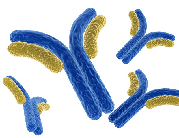
The investigative team utilized an antibody designated YN9303-24, which promotes the open conformation of TMEM63B. Through chimeric proteins, progressively shortened C-terminal truncations, and targeted amino acid deletions, the researchers mapped the antibody-binding site to the intracellular C-terminal tail. Within this region, a three-amino-acid sequence known as the AQV motif at positions 773–775 was identified as the primary antibody recognition site.
Adjacent to this epitope, the LQD sequence spanning residues 776–778 was found to directly control TMEM63B activity. Eliminating these three amino acids produced a constitutively active protein capable of executing lipid scrambling without membrane stimulation. Additional targeted substitutions isolated Leucine at 776 as the principal modulatory residue. Replacing Leu776 with alanine substantially increased the exposure of phosphatidylserine (PS) on the cell surface—a phospholipid normally confined to the inner cytoplasmic leaflet under resting conditions. Enhanced lipid scrambling was further confirmed via increased uptake of fluorescent phosphatidylcholine.
“The C-terminal tail functions like a molecular brake, keeping TMEM63B inactive under resting conditions until changes in the membrane allow it to become activated,” explains Professor Katsumori Segawa of the Department of Medical Chemistry at the Institute of Science Tokyo.
Structural Interactions Within the Intracellular Domain
Further structural analysis clarified the operational mechanics of this molecular brake. Within the open-state structure, the AQVLQD motif positions directly adjacent to two intracellular helices, designated IL2H2 and IL2H3, which construct a beam-like intracellular domain of TMEM63B. Proximity between Leu776 and multiple hydrophobic residues across these helices indicates that hydrophobic interactions between the C-terminal tail and the intracellular domain stabilize the closed, inactive conformation. Disrupting these interactions releases the structural restraint, permitting the lipid translocation pathway to open.
These molecular insights carry direct relevance for neuropathology. Earlier studies have linked disease-associated mutations within TMEM63B to neurodevelopmental and neurodegenerative disorders, suggesting that failure of this autoinhibitory brake may trigger pathological cellular signaling.