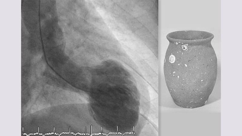
Takotsubo Cardiomyopathy Triggers Rare Coronary Embolism
A 74-year-old woman presenting with acute chest pain and shortness of breath two days after laser cataract surgery was found to have a rare coronary embolism linked to takotsubo cardiomyopathy and a left ventricular mural thrombus.
Key Clinical Takeaways:
- A 74-year-old woman developed an acute coronary embolism following laser cataract surgery, during which her apixaban was withheld for three days.
- Emergency diagnostic evaluations revealed ST-elevation myocardial infarction patterns alongside classic imaging features of takotsubo cardiomyopathy and left ventricular mural thrombus.
- Medical personnel successfully performed a balloon angioplasty and thrombectomy on the distal obtuse marginal 1 branch without encountering atherosclerotic obstructive coronary artery disease.
Patient Reports Chest Pain After Pausing Apixaban
The patient arrived at the emergency department reporting persistent chest pain and shortness of breath lasting two days. Her medical history included chronic obstructive pulmonary disease, tobacco use disorder, dyslipidemia, generalized anxiety disorder, and a prior unprovoked deep vein thrombosis and pulmonary embolism managed with apixaban. Two days prior to her emergency admission, she underwent laser cataract surgery, which required pausing her apixaban therapy for three days.
Initial vital signs and physical examinations yielded normal ranges. However, an electrocardiogram displayed ST elevation and pathologic Q waves located in the inferior and anterolateral leads, pointing toward an ST-elevation myocardial infarction. Laboratory results recorded an initial troponin level of 5827 ng/L against a normal range under 14 ng/L, while B-type natriuretic peptide reached 10,861 pg/ln compared to a normal range below 100 pg/mL.
Diagnostic Imaging and Catheterization Findings
Cardiac catheterization identified an embolus featuring two truncations within the distal obtuse marginal 1 branch. The procedure involved administering a loading dose of tirofiban and clopidogrel alongside a single intravenous dose of furosemide. Medical staff performed balloon angioplasty and thrombectomy, successfully clearing the obstruction in the distal obtuse marginal 1 branch without any observed atherosclerotic obstructive coronary artery disease.
Transthoracic echocardiography displayed a classic takotsubo cardiomyopathy presentation defined by basal hyperdynamic movement, apical and anterior wall ballooning, and the presence of a left ventricular mural thrombus. A computed tomography pulmonary angiogram ruled out pulmonary embolism. Apixaban treatment resumed within 24 hours following the procedure.
Medical Team Prescribes Dual Antiplatelet Therapy
In their detailed report of the case, Boran Mao, MD, and colleagues from the Internal Medicine department at the University of Central Florida College of Medicine in Orlando described the presentation as an unusual, multifaceted coronary embolism caused by takotsubo cardiomyopathy.
The medical team discharged the patient on a comprehensive pharmacological and device regimen. Her prescriptions included dual antiplatelet therapy consisting of 81 mg of aspirin once daily and 75 mg of clopidogrel once daily, alongside apixaban at 5 mg twice daily. Guideline-directed medical therapy featured valsartan at 24 mg twice daily, sacubitril at 26 mg twice daily, and carvedilol at 6.25 mg twice daily. She also received a wearable defibrillator vest prior to leaving the facility.
When discussing how to handle these situations, the researchers highlighted that managing very high-risk patients to stop potentially fatal thromboembolic episodes necessitates collaborative input from a multidisciplinary group.