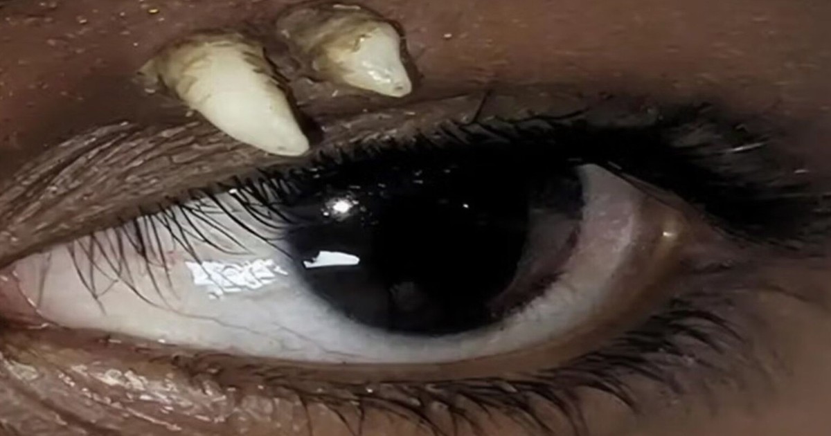
Rare Case: Fully Developed Tooth Found in 16-Year-Old Boy’s Eye Socket
A completely developed permanent canine tooth was discovered inside the upper orbital cavity of a 16-year-old boy in Madurai, India, according to a medical case report published in June 2026 by the American Academy of Ophthalmology and detailed by Egészségkalauz. The rare anatomical anomaly was identified after the teenager sought medical evaluation for a persistent, painless swelling on his upper left eyelid that had developed approximately one month prior with no history of trauma.
Initial Medical Examination at Aravind Eye Hospital
Specialists at the Aravind Eye Hospital in Madurai first detected two hard, whitish, well-defined masses near the eyebrow during physical examinations of the patient’s upper left eyelid. To investigate the unusual presentation, clinicians ordered a computed tomography (CT) scan. The imaging revealed a structure with a clearly formed crown and root located in the upper part of the left eye socket, protruding outward toward the eyelid.
Surgeons subsequently performed an operation to remove the hard structure, confirming during the procedure that it was a fully formed canine tooth. Because ectopic teeth can occasionally mimic neoplasms, the medical team tested the extracted tissue for teratoma—a type of tumor capable of producing diverse tissues such as hair, bone, or tooth-like structures. Diagnostic evaluations ruled out teratoma, leading physicians to diagnose the condition as an ectopic tooth, meaning a tooth that develops outside the dental arch.
Embryonic Disruptions Cause Ectopic Tooth Formation
Teeth occasionally develop far from their normal locations within the oral cavity due to disruptions during early embryonic development. The formation of the face, jaws, and dentition relies on precise interactions between various cell groups and tissues at specific times. When this complex process encounters an interruption, a tooth germ can migrate or develop entirely outside the mouth, sometimes appearing in the nasal cavity, maxillary sinus, palate, or orbit.
In addition to developmental anomalies during gestation, ectopic teeth in the upper jaw can be triggered by trauma, previous dental procedures, cysts, or tumors, as outlined in a review examining ectopic teeth in the maxilla. Genetic factors also contribute when extra, or supernumerary, teeth develop, a characteristic seen in rare hereditary conditions such as cleidocranial dysplasia. However, clinicians emphasize that a single ectopic tooth does not automatically indicate an underlying genetic syndrome.
Review of Literature and World Rarity
Dr. Meghana Tanwar, one of the authors of the case study, noted that a review of medical literature yielded no prior documented reports of a fully developed tooth discovered in the upper region of the eye socket in a position identical to the Indian teenager’s case. While ectopic teeth have been documented in the maxillary sinus and nasal cavity, an orbital location remains exceptionally rare.
Symptoms associated with ectopic teeth depend entirely on their exact anatomical location and the surrounding tissues involved. Teeth lodged in the nasal cavity frequently cause chronic nasal congestion, discharge, or recurrent nosebleeds, whereas orbital presentations can manifest primarily as localized swelling. Diagnostic confirmation heavily relies on CT imaging, which provides detailed visualization of bony structures relative to adjacent nerves, blood vessels, and ocular elements.
Treatment Depends on Tooth Location and Symptoms
Treatment decisions for ectopic teeth depend on symptom severity, location, and the risk of complications. Asymptomatic cases that pose no threat to surrounding tissues may simply require regular clinical monitoring. Conversely, intervention becomes necessary when an ectopic tooth causes infection, pain, or structural pressure.
Surgical approaches vary by location, with nasal or sinus cases frequently managed via endoscopic techniques where surgeons operate through the nasal passages without external incisions, supported by findings from a literature review analyzing 27 cases. Following the removal of the ectopic tooth from the orbital cavity, the patient in Madurai showed no signs of additional supernumerary teeth, and medical staff assessed the likelihood of recurrence as very low.