Patient Develops QT Prolongation and Torsades de Pointes After Pulsed Field Ablation
A patient in his 70s developed marked QT prolongation and torsades de pointes following pulsed field ablation for persistent atrial fibrillation. The event occurred alongside severe ventricular dysfunction, residual amiodarone effects, and postoperative hypokalemia.
- A man in his 70s experienced torsades de pointes on postoperative day two after undergoing pulsed field ablation for persistent atrial fibrillation.
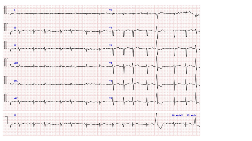
- Post-procedure evaluation revealed a corrected QT interval reaching 610 milliseconds on postoperative day one.
- Clinical documentation highlights that multifactorial elements—including underlying heart disease, prior amiodarone exposure, and electrolyte imbalances—prevent direct attribution of the arrhythmia to pulsed field ablation alone.
Patient Presents with Heart Failure and Atrial Fibrillation
The patient was admitted in 2025 presenting with persistent atrial fibrillation and chronic heart failure. His prior medical record included impaired glucose tolerance, premature ventricular contractions, and an earlier hospitalization for heart failure roughly a decade prior. Approximately three months before the procedure, an asymptomatic 14-beat episode of nonsustained ventricular tachycardia prompted the initiation of amiodarone at 200 milligrams daily. Transthoracic echocardiography documented a left ventricular ejection fraction of 18%, while baseline automated electrocardiographic readings showed a corrected QT interval of 395 milliseconds. Coronary angiography confirmed the absence of significant stenosis.
Pulmonary vein isolation was performed using the FARAPULSE system manufactured by Boston Scientific Corporation, operating under intravenous anesthesia with propofol and rocuronium. The medical team delivered 40 total applications across all four pulmonary veins, confirming electrical isolation before concluding the procedure with one 10-joule intracardiac cardioversion. Amiodarone treatment was discontinued on the day of the procedure. Recovery featured a brief period of delayed awakening and shallow respiration requiring four hours of noninvasive positive-pressure ventilation, alongside a single intravenous dose of 20 milligrams of furosemide administered for suspected mild right-sided heart failure.
Postoperative QT Prolongation Triggers Torsades De Pointes
One hour after the procedure, an electrocardiogram recorded a sinus rhythm with a corrected QT interval of 431 milliseconds. By postoperative day one, however, monitoring captured a marked prolongation of the corrected QT interval to 610 milliseconds alongside frequent premature ventricular contractions. On postoperative day two, telemetry recorded a short-long-short sequence initiated by a premature ventricular contraction that immediately preceded a bout of torsades de pointes. The episode induced altered consciousness and required a single 150-joule direct-current shock to terminate.
Medical staff initiated electrolyte repletion following the arrhythmic event. Intravenous potassium chloride was administered in 20-mEq increments to maintain serum potassium above 4.0 mEq/L, and a single 20-mEq dose of magnesium sulfate was given. No further episodes of torsades de pointes occurred. By postoperative day four, the corrected QT interval shortened to 479 milliseconds. At a one-month follow-up assessment, the corrected QT interval returned to 403 milliseconds, and the patient’s left ventricular ejection fraction improved to 30%, climbing further to 36% at six months.
The evaluation of abnormal ventricular repolarization extends beyond procedural complications. Drug-induced QT prolongation represents a known risk factor for early after-depolarizations and subsequent re-entrant tachycardias like torsades de pointes. A corrected QT interval exceeding 500 milliseconds or an acute increase greater than 60 milliseconds generally indicates a high risk for malignant ventricular arrhythmias. While Class I and Class III anti-arrhythmic medications are established contributors, non-cardiac drugs such as specific anti-infectives, antiemetics, and antipsychotics also carry recognized potential to alter myocardial repolarization when secondary risk factors are present.
The patient’s severe ventricular dysfunction, residual electrophysiological effects from amiodarone exposure, and postoperative hypokalemia created a fragile arrhythmogenic substrate. While conventional pulmonary vein isolation has been attributed, at least in part, to modification of the cardiac autonomic nervous system and rare cases of torsades de pointes, the precise electrophysiological contribution of pulsed field ablation remains incompletely understood.
Disclaimer: The information provided in this article is for educational and scientific communication purposes only and does not constitute medical advice. Always consult with a qualified healthcare provider regarding any medical condition, diagnosis, or treatment plan.
Worth a look