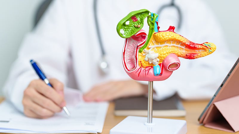
Newer EUS Needle Designs Improve Diagnostic Accuracy for Autoimmune Pancreatitis
Endoscopic ultrasound-guided biopsy techniques are improving diagnostic accuracy for autoimmune pancreatitis, addressing a long-standing clinical gap where imaging and serology frequently fall short. According to clinical data published on Pancreapedia and Medscape Medical News, specialized needle designs and tissue acquisition methods allow clinicians to secure histological samples from a condition that mimics pancreatic malignancies.
- Endoscopic ultrasound-guided tissue acquisition helps differentiate autoimmune pancreatitis from pancreatic cancer when standard imaging is inconclusive.
- Newer needle designs outperform older models in acquiring diagnostic core samples from the affected pancreatic parenchyma.
- Accurate diagnosis via these techniques reduces diagnostic delays and prevents unnecessary surgical interventions or risky steroid trials.
The Diagnostic Dilemma in Autoimmune Pancreatitis
Autoimmune pancreatitis has historically presented major diagnostic hurdles. The condition frequently manifests as a focal solitary mass in the head of the pancreas, causing obstructive jaundice and upstream duct dilation. These features mimic locally advanced pancreatic cancer, often leading to inappropriate surgical resections or protracted diagnostic steroid trials that carry inherent patient safety risks. Current international consensus diagnostic criteria incorporate five cardinal features—imaging characteristics of the pancreas, serology, other organ involvement, pancreatic histology, and response to steroids—yet these measures leave many cases clinically ambiguous.
Evolving Utility of Endoscopic Ultrasound Imaging and Biopsy
While standard computed tomography and magnetic resonance cholangiopancreatography provide baseline structural views, they cannot definitively rule out malignancy without tissue confirmation. Endoscopic ultrasound bridges this gap by offering high-resolution imaging of the pancreatic parenchyma alongside direct tissue acquisition via fine-needle aspiration or trucut biopsy. Data from Pancreapedia show that standard endoscopic ultrasound reveals a diffusely enlarged gland with patchy, heterogeneous parenchyma in up to 57 percent of patients with classic features. However, because many patients present with atypical findings or localized masses, imaging alone remains insufficient without histological validation.
Refining Needle Technology for Enhanced Histology
Obtaining adequate core tissue samples for histological analysis remains vital for confirming autoimmune pancreatitis. Newer endoscopic ultrasound needle designs yield superior results compared to older models. By securing higher-quality core samples, medical teams can bypass invasive exploratory surgeries and establish targeted therapy earlier in the disease course.
*Disclaimer: The information provided in this article is for educational and scientific communication purposes only and does not constitute medical advice. Always consult with a qualified healthcare provider regarding any medical condition, diagnosis, or treatment plan.*