New Genetic Sensor Enables MRI to Image Molecular-Level Changes
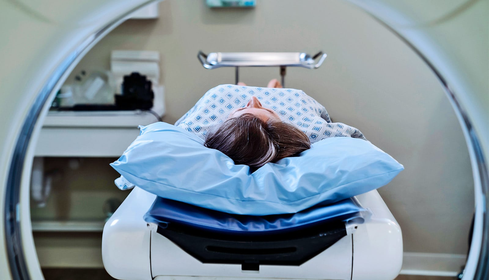
Most patients encounter magnetic resonance imaging (MRI) as a noisy, claustrophobic necessity—a diagnostic tool that provides a high-resolution map of the body’s internal architecture. However, in the clinical world, architecture is often a lagging indicator. By the time a tumor alters the morphology of an organ or a neurodegenerative disease erodes brain tissue, the window for early intervention has frequently closed.
Key Clinical Takeaways:
- Researchers have developed MAPPER, a genetically encoded protein-based sensor that allows MRIs to visualize molecular-level cellular activity in real-time.
- The system utilizes aquaporin proteins to manipulate water molecule movement, creating a magnetic signal that reveals chemical processes before structural damage occurs.
- This modular “LEGO-like” architecture can be tailored to detect multiple different analytes, potentially transforming the study of cancer metastasis and neurodegeneration.
The fundamental limitation of traditional MRI has always been its reliance on anatomical changes. Whereas the technology is peerless in visualizing the brain, heart, and kidneys without the risks associated with ionizing radiation, it essentially captures a snapshot of the “house” rather than the “activity” inside the rooms. For clinicians, this creates a dangerous diagnostic gap: the pathogenesis of a disease is often well underway at the molecular level long before it manifests as a visible lesion or tissue atrophy on a scan.
The Biological Mechanism of MAPPER
To bridge this gap, researchers at the University of California, Santa Barbara, have introduced a system known as MAPPER (modular aquaporin-based protease-activatable probes for enhanced reporting). This innovation shifts the focus from macro-structures to the behavior of water molecules within the cell. As detailed in the peer-reviewed journal Science Advances, the system leverages aquaporin—a protein that forms hourglass-shaped channels in the cell membrane to regulate water flow.

Because water molecules function as tiny magnets, controlling the rate at which they move across the cell membrane allows the MRI to pick up a specific magnetic signal. This signal is not based on the presence of a mass, but on a biological process. By genetically engineering these sensors into cells, the MRI can “observe” molecular activity as it happens. This approach mirrors the use of jellyfish-derived fluorescent proteins in microscopy, but translates that precision to the deep-tissue capabilities of the MRI.
“If we can see these molecular-level changes happening in real time, then we can ask questions like, ‘How do tumor cells metastasize?’ or ‘How does neurodegeneration progress at the molecular level as an animal ages?'”
— Arnab Mukherjee, Associate Professor of Chemical Engineering, UCSB
The modularity of the MAPPER system is its most significant clinical advantage. Rather than designing a new sensor for every unique protein or chemical, researchers can substitute specific proteins into the architecture to target different cellular processes. This flexibility allows the system to detect nearly ten different systems within a single setup, a stark contrast to previous genetic sensors that were typically limited to a single analyte.
Clinical Implications for Neurodegeneration and Oncology
The ability to track molecular changes in real-time has profound implications for the standard of care in oncology and neurology. In current practice, determining if a drug is successfully arresting the growth of cancer cells often requires waiting for the tumor to shrink—a structural change. With a molecular sensor, clinicians could potentially observe the biochemical failure of cancer cells long before the tumor’s physical dimensions change.
Similarly, in the realm of neurodegenerative diseases, the progression of the disease is often invisible until significant cognitive decline occurs. The MAPPER system could allow researchers to monitor calcium changes in the brain or the early biochemical markers of protein misfolding, providing a window into the disease’s progression in its earliest stages. For patients currently navigating these diagnoses, maintaining a rigorous relationship with board-certified neurologists is critical to utilize the most advanced current diagnostic protocols while these molecular tools move toward human application.
This movement toward “MRI-plus” technologies is a broader trend in radiology. For instance, the MRI Research Institute (MRIRI) at Weill Cornell Medicine focuses on similar goals, developing quantitative methods and data science techniques to improve disease detection and precision medicine. The integration of molecular sensors like MAPPER into these existing research frameworks could accelerate the translation of this technology from the benchtop to the bedside.
Transforming Preclinical Research and Animal Welfare
Beyond direct human application, the MAPPER system addresses a critical ethical and scientific hurdle in animal studies. Currently, studying the internal organs of a living subject often requires “sacrificing” the animal to obtain a physical sample, providing only a single snapshot in time. This method is frequently misleading due to variations in metabolism and individual responses to treatment across different subjects.
“Our approach allows continuous imaging of the same animal over the course of a study, giving a far more accurate picture of disease and of biology.”
— Asish Ninan Chacko, PhD researcher
By enabling longitudinal imaging of the same subject, the MAPPER system reduces the number of animals required for studies and increases the statistical reliability of the data. This shift toward continuous monitoring allows for a more nuanced understanding of how a disease evolves, potentially shortening the timeline for drug development and safety testing.
Navigating the Transition to Molecular Diagnostics
While the MAPPER system represents a leap forward, the path to widespread clinical adoption involves significant regulatory and technical hurdles. Integrating genetically encoded sensors into human medicine requires rigorous safety profiles and FDA oversight. In the interim, the focus remains on refining these tools in laboratory settings and animal models to ensure absolute specificity and sensitivity.
For healthcare providers and diagnostic centers, the evolution of MRI technology necessitates a constant update of imaging protocols. Facilities that prioritize the latest in diagnostic imaging services are best positioned to integrate these advancements as they emerge from the research phase. The goal is a future where an MRI does not just show us what the body looks like, but how it is functioning at the most fundamental level.
As we move toward an era of truly precision medicine, the ability to detect a disease before it alters anatomy will be the defining characteristic of early intervention. The work coming out of UCSB, supported by the broader academic community and institutions like Columbia University’s Center for MRI Research, suggests that the “silent” phase of many diseases may soon become visible.
Looking ahead, the trajectory of medical imaging is clear: the transition from anatomy to activity. By turning water molecules into reporters of cellular health, we are moving closer to a world where “too late” is no longer the standard answer for many chronic conditions. To ensure you are receiving the highest standard of current care, we encourage patients to utilize our directory to find vetted diagnostic centers and specialists dedicated to the latest evidence-based practices.
Disclaimer: The information provided in this article is for educational and scientific communication purposes only and does not constitute medical advice. Always consult with a qualified healthcare provider regarding any medical condition, diagnosis, or treatment plan.