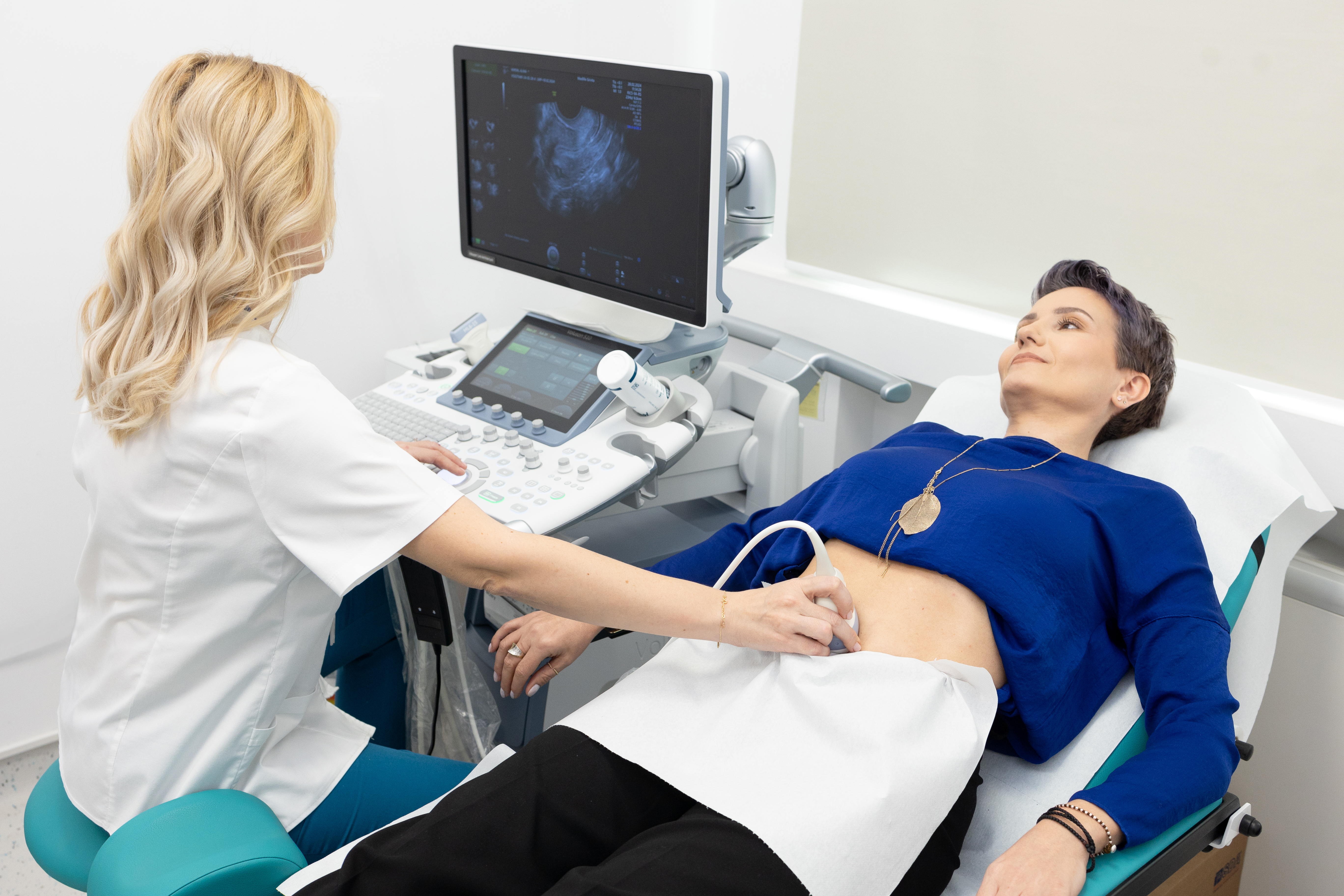
Medical Tests and Ultrasound for Intermenstrual Bleeding Causes
Unscheduled uterine bleeding outside of regular menstruation requires immediate clinical evaluation to rule out structural pathologies, hormonal dysfunctions, or malignant neoplasms, with transvaginal ultrasound serving as a primary diagnostic instrument. According to clinical guidelines and health reporting from HotNews.ro, atypical spotting or hemorrhage demands systematic investigation to identify underlying etiologies ranging from benign endometrial polyps to malignant transformations.
Key Clinical Takeaways:
- Transvaginal ultrasound provides high-resolution visualization of the myometrium and endometrium, making it an indispensable first-line tool for identifying structural anomalies like fibroids and polyps.
- Persistent intermenstrual bleeding requires a comprehensive diagnostic panel, including complete blood counts, hormonal assays, and targeted imaging.
- Timely specialized consultation through a certified gynecology clinic ensures rapid risk stratification and prevents the progression of untreated conditions.
The Diagnostic Imperative of Transvaginal Ultrasound
When a patient presents with abnormal uterine bleeding, establishing an accurate anatomical baseline is the immediate priority for attending physicians. Transvaginal ultrasonography allows clinicians to measure endometrial thickness, assess myometrial echogenicity, and detect focal lesions such as submucosal myomas or endometrial hyperplasia with high sensitivity. Per data highlighted in clinical overviews published via PubMed, pelvic imaging remains the non-invasive standard of care for differentiating between organic lesions and dysfunctional uterine bleeding. Identifying these structural anomalies early alters the therapeutic trajectory, sparing patients from prolonged morbidity.
Laboratory Investigations and Hormonal Profiling
Imaging alone rarely paints a complete clinical picture. Standard diagnostic protocols mandate a rigorous battery of blood tests to evaluate systemic contributors to hemorrhage. Physicians routinely order a complete blood count to screen for iron-deficiency anemia resulting from chronic blood loss, alongside thyroid-stimulating hormone (TSH) assays and prolactin levels to uncover endocrine disruptions that frequently trigger ovulatory dysfunction. Furthermore, evaluating coagulation profiles helps rule out underlying bleeding disorders, ensuring that therapeutic interventions address both symptom management and primary pathogenesis. For personalized diagnostic pathways, patients should consult with an experienced endocrinologist or gynecological specialist.
Advanced Histopathological Evaluation and Hysteroscopy
When non-invasive imaging reveals endometrial thickening or persistent focal abnormalities, definitive diagnosis requires direct visualization and tissue acquisition. Hysteroscopy combined with targeted biopsy represents the gold standard for histological verification, allowing pathologists to examine cellular architecture for atypical hyperplasia or carcinoma. According to clinical standards tracked by major health agencies such as the World Health Organization, early histological confirmation of atypical bleeding patterns significantly improves prognosis for malignant diagnoses. Coordinating these multi-step interventions smoothly often requires professional clinical guidance, making it valuable to connect with a specialized diagnostic healthcare center equipped for comprehensive ambulatory care.
*Disclaimer: The information provided in this article is for educational and scientific communication purposes only and does not constitute medical advice. Always consult with a qualified healthcare provider regarding any medical condition, diagnosis, or treatment plan.*