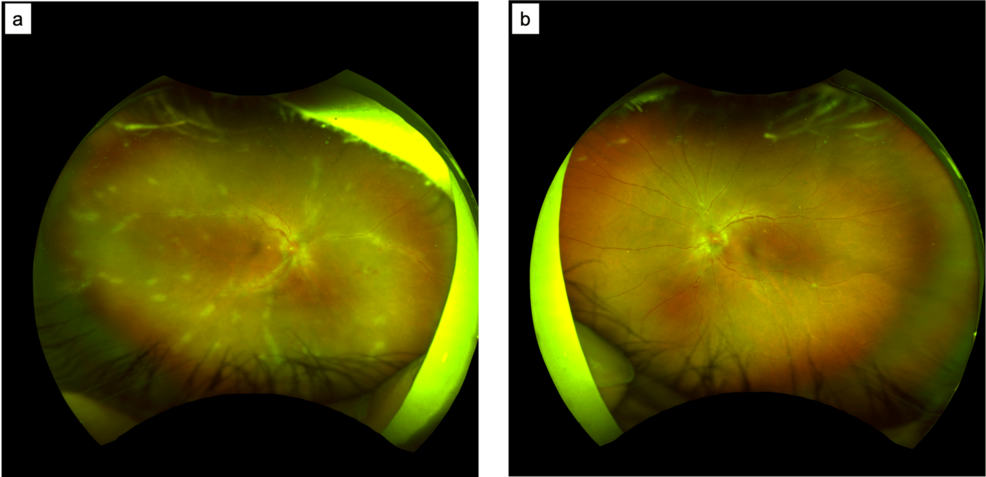
HTLV-1 Associated Uveitis and Frosted Retinal Vasculitis
Human T-lymphotropic virus type 1 (HTLV-1)-associated uveitis presenting with frosted retinal vasculitis represents a complex diagnostic challenge at the intersection of retrovirology and ophthalmology, according to clinical findings published in Cureus. As clinicians increasingly encounter this condition in non-endemic regions due to migration patterns documented by EyeWiki, understanding the precise mechanisms of intraocular inflammation is critical for effective patient management.
- HTLV-1-associated uveitis can present as the sole manifestation of this retroviral infection, featuring vitreous opacities and severe retinal vasculitis.
- Polymerase chain reaction analysis frequently detects HTLV-1 proviral DNA in the aqueous humor of affected patients, confirming an immune-mediated mechanism driven by infected CD4+ T cells.
- Managing complex ocular inflammation requires prompt coordination with vetted board-certified ophthalmologists and specialized diagnostic centers.
HTLV-1 is an oncovirus first isolated in 1980 by Poiesz and colleagues, with approximately 20 million individuals estimated to be infected worldwide, per data compiled on EyeWiki. While the majority of carriers remain asymptomatic, the virus is endemic to specific geographic regions including Japan, Melanesia, the Caribbean Islands, Central and South America, and central African areas. Ocular manifestations such as uveitis occur in roughly 112.2 per 100,000 HTLV-1 carriers of both sexes. Transmission primarily occurs vertically through prolonged breastfeeding, via blood contact, or through sexual intercourse, with prevalence increasing notably among individuals over 40 years of age, particularly women.
The pathogenesis of HTLV-1 uveitis stems from an immune-mediated response within the eye. Analysis of aqueous humor samples reveals that floating cells in the anterior chamber consist predominantly of CD3+ T lymphocytes and macrophages. Polymerase chain reaction analysis detects HTLV-1 proviral DNA, viral mRNA, and viral proteins within the aqueous humor of these patients, but notably not in patients with uveitis of alternative etiologies. This localized presence of infected CD4+ T cell clones underscores a distinct immunopathogenic profile that differentiates HTLV-1 uveitis from idiopathic intraocular inflammation.
Historically linked to adult T-cell leukemia/lymphoma and HTLV-1-associated myelopathy/tropical spastic paraparesis, the virus also targets ocular tissues through malignant infiltrates, corneal damage, retinal degeneration, and neurophthalmic disorders. When patients present with atypical intraocular inflammation, ruling out underlying retroviral pathology is essential.
Accurate diagnosis relies on recognizing the distinct clinical overlap between viral infection and autoimmune presentation. Because HTLV-1 uveitis frequently mimics other forms of posterior or intermediate uveitis, misdiagnosis can lead to inappropriate therapeutic regimens.
As epidemiological shifts continue to introduce vector-associated pathologies into non-endemic zones, maintaining a high index of suspicion remains paramount.
*Disclaimer: The information provided in this article is for educational and scientific communication purposes only and does not constitute medical advice. Always consult with a qualified healthcare provider regarding any medical condition, diagnosis, or treatment plan.*