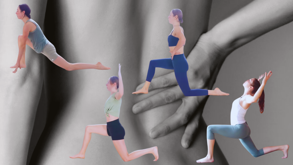
How to Practice Low Lunge Without Straining Your Low Back
Low lunge, or Anjaneyasana, is a foundational yoga posture frequently associated with lumbar strain when performed with improper pelvic alignment or insufficient core engagement. Clinical observation suggests that the primary mechanism of injury involves excessive anterior pelvic tilt, which increases compressive forces on the facet joints of the lumbar spine. According to a systematic review published in the Journal of Bodywork and Movement Therapies, spinal morbidity in yoga practitioners is often correlated with repetitive hyperextension that exceeds the individual’s functional range of motion.
Key Clinical Takeaways:
- Lumbar strain in low lunge is typically caused by excessive anterior pelvic tilt rather than the lunge depth itself.
- Engaging the deep stabilizers—specifically the transverse abdominis and gluteus maximus—is essential to neutralize spinal compression.
- Persistent mechanical lower back pain should be evaluated by a professional to rule out spondylolysis or disc-related pathology.
Biomechanical Pathogenesis of Lumbar Compression
The lumbar spine is designed to support load in a neutral alignment. When a practitioner enters a low lunge without stabilizing the pelvis, the psoas muscle—which attaches to the lumbar vertebrae—can pull the spine into hyperlordosis. Dr. Elena Rodriguez, a specialist in orthopedic sports medicine, notes that “the structural integrity of the L4-L5 segment is highly vulnerable to shear forces when the pelvis tilts forward, effectively shortening the posterior space of the spinal canal.”
Research funded by the National Institutes of Health (NIH) on musculoskeletal kinetics indicates that force distribution is optimized when the pelvic floor is aligned beneath the ribcage. Failure to maintain this alignment forces the lumbar extensors to compensate, leading to localized myofascial fatigue and potential chronic inflammation. For individuals experiencing recurring discomfort, it is critical to seek a formal assessment. Patients requiring diagnostic imaging or gait analysis should consider consulting with a board-certified physical therapist or sports medicine specialist to identify underlying biomechanical asymmetries.
Corrective Strategies and Clinical Standard of Care
To mitigate the risk of injury, the current clinical standard of care emphasizes “posterior pelvic tilt” as a protective mechanism. By actively engaging the gluteus maximus of the trailing leg, the practitioner creates reciprocal inhibition of the hip flexors, which reduces the pull on the lumbar spine. This approach is supported by longitudinal data regarding preventative biomechanical training, which suggests that neuromuscular re-education can significantly decrease the incidence of training-related injuries.
Beyond engagement, the depth of the lunge must be titrated to the practitioner’s specific hip mobility. “Pushing into a range of motion that exceeds the hip capsule’s capacity will inevitably force the lumbar spine to ‘borrow’ mobility,” says Dr. Marcus Thorne, a PhD researcher in kinesiology. “This is a primary driver of non-traumatic lumbar strain in clinical settings.” For those who have already sustained soft tissue injury, access to specialized rehabilitative care is paramount. We recommend utilizing our directory to connect with a vetted orthopedic rehabilitation center to ensure that any corrective exercise program is tailored to your specific diagnostic profile.
Evaluating the Risk of Chronic Morbidity
While yoga is widely recognized for its therapeutic benefits, it is not without contraindications. The prevalence of “yoga-induced” back pain often arises from a misunderstanding of spinal loading. A report in the British Medical Journal (BMJ) highlights that while adverse events are relatively rare, they are almost exclusively linked to improper technique rather than the inherent nature of the postures themselves.

For practitioners, the diagnostic gap remains a significant barrier to safe practice. If you are experiencing radiating pain, numbness, or weakness—indicators of potential radiculopathy—do not attempt to self-correct through further stretching. Clinical triage is necessary to rule out herniated discs or nerve impingement. We advise patients to reach out to a diagnostic imaging facility or spine specialist to obtain a definitive diagnosis before resuming high-intensity movement practices.
Future Trajectories in Movement Science
The evolution of movement science is moving toward highly individualized, data-driven protocols. Future research will likely focus on real-time kinematic feedback systems that allow practitioners to monitor pelvic tilt during static holds. As we await more granular data on the long-term efficacy of these interventions, the emphasis remains on conservative management and professional supervision. Maintaining optimal spinal health requires a proactive approach to biomechanics and a willingness to prioritize anatomical safety over aesthetic depth.
Disclaimer: The information provided in this article is for educational and scientific communication purposes only and does not constitute medical advice. Always consult with a qualified healthcare provider regarding any medical condition, diagnosis, or treatment plan.