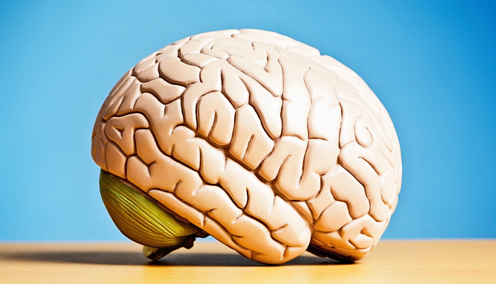
How Brains Clear Harmful Waste Using the Glymphatic System
Artificial intelligence is bridging a critical gap in neurobiology by enabling the high-resolution mapping of the glymphatic system—the brain’s specialized waste-clearance pathway. By integrating physics-informed machine learning with standard magnetic resonance imaging (MRI), researchers have successfully quantified the velocity of cerebrospinal fluid (CSF) flow, providing a potential diagnostic window into the pathogenesis of neurodegenerative conditions like Alzheimer’s disease.
Key Clinical Takeaways:
- Quantifying Clearance: New AI-driven models allow clinicians to measure the speed of fluid flow in the brain, distinguishing between rapid surface circulation and slower, deep-tissue perfusion.
- Diagnostic Potential: This technology could eventually serve as a biomarker for early-stage Alzheimer’s, identifying “poor circulation” in the brain long before significant cognitive decline manifests.
- Broad Applications: Beyond dementia, this methodology offers a pathway to monitor recovery from traumatic brain injuries (TBI) by assessing the structural integrity of the brain’s waste-removal systems.
The Mechanics of Metabolic Clearance
The glymphatic system, first characterized by Dr. Maiken Nedergaard in a 2012 study published in Science, functions as a macroscopic waste-disposal network. During deep, non-REM sleep, the brain facilitates the exchange of interstitial fluid and CSF to flush out metabolic byproducts, including amyloid-beta and tau proteins—the primary constituents of the plaques and tangles associated with Alzheimer’s. Despite the established biological importance of this system, clinical assessment has remained elusive due to the technical limitations of traditional neuroimaging.

“MRI is an indispensable tool for non-invasive structural imaging, yet it has historically struggled to capture the ultra-slow fluid dynamics occurring within the brain’s parenchyma,” explains Dr. Douglas Kelley of the University of Rochester. By applying physics-informed neural networks to MRI data, the research team—whose work was recently published in Science Advances—has overcome these limitations. The study, supported by the National Institutes of Health (NIH) and the NIH BRAIN Initiative, demonstrates that fluid flow is not uniform; it operates at distinct velocities depending on the region of the central nervous system.
Clinical Triage and the Future of Neuro-Imaging
The ability to map these flow velocities in vivo creates a new paradigm for specialized neuro-diagnostic centers, which may soon be able to incorporate glymphatic mapping into standard cognitive health screenings. For patients presenting with early-onset cognitive symptoms or those with a high genetic predisposition for Alzheimer’s, early detection of impaired glymphatic clearance could shift the focus from reactive treatment to proactive, lifestyle-based neuro-protection.
If you or a family member are navigating the complexities of memory disorders, it is essential to consult with board-certified neurologists who specialize in advanced imaging interpretation and neurodegenerative disease management. Early intervention, guided by emerging biomarkers, remains the most effective strategy for mitigating long-term neurological morbidity.
Addressing the Challenges of Neuro-Degeneration
The research reveals that the glymphatic system utilizes two distinct flow patterns. Surface regions exhibit fluid movement of a few microns per second, while deep-tissue clearance occurs at a rate approximately 50 times slower. This kinetic disparity suggests that the brain’s vulnerability to protein accumulation is not uniform, but rather dependent on the localized permeability of the tissue.

“We are moving from a state of knowing that the brain cleans itself to a state of being able to measure exactly how efficiently that process is occurring in a living patient,” notes Dr. Elena Rossi, an independent neuro-researcher not affiliated with the study. “This is the difference between diagnosing a systemic failure and pinpointing a specific mechanical blockage.”
For clinical organizations and healthcare compliance entities, the integration of AI-driven diagnostic tools necessitates a rigorous review of current data-processing protocols. As these algorithms transition from animal models to human clinical trials, health systems must ensure that their diagnostic infrastructure is equipped to handle the high-dimensional data required for glymphatic mapping.
Establishing Baseline Data for Future Trials
The researchers are currently refining their AI models using murine baseline data. The goal is to transition toward human longitudinal studies, where the technology can be used to compare healthy aging brains with those exhibiting signs of neurodegeneration. This transition is critical for validating the standard of care for dementia prevention. By identifying those with compromised fluid circulation, clinicians may eventually be able to intervene with therapeutic strategies designed to enhance glymphatic function, such as sleep optimization or pharmacological agents that influence CSF pressure.
As we advance toward these human-centric applications, the collaboration between mechanical engineering, neurobiology, and clinical neurology will define the next decade of geriatric care. Patients and providers alike should monitor the progress of these Phase I-equivalent data-modeling studies as they move closer to clinical validation.
Disclaimer: The information provided in this article is for educational and scientific communication purposes only and does not constitute medical advice. Always consult with a qualified healthcare provider regarding any medical condition, diagnosis, or treatment plan.