Furuncular Cutaneous Myiasis in a Nontraveler: A Case Report
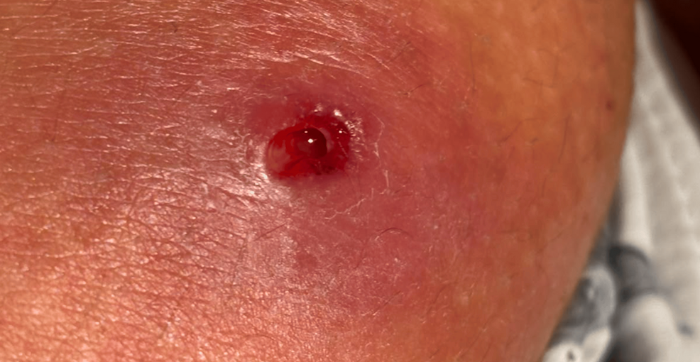
Furuncular cutaneous myiasis, a parasitic infestation where fly larvae embed in the skin, can occur in individuals who have not traveled to endemic regions, as documented in a clinical case report published in Cureus. This condition typically presents as a painful, boil-like lesion with a central pore that may exude fluid or show larval movement, requiring precise surgical extraction for resolution.
- Clinical Presentation: Appears as a furuncle (boil) with a characteristic “punctum” or opening where the larva breathes.
- Diagnostic Challenge: Often misdiagnosed as a bacterial abscess or cyst due to the absence of a travel history.
- Standard Treatment: Mechanical removal of the larva, sometimes aided by occlusion to force the parasite to the surface.
The case reported in Cureus highlights a critical diagnostic gap: the assumption that cutaneous myiasis is exclusively a “traveler’s disease.” The patient presented with a lesion that mimicked a common skin abscess, leading to initial diagnostic uncertainty. This pathogenesis occurs when dipterous flies deposit eggs or larvae on the skin, or when a secondary vector, such as a mosquito, transports the larvae to the human host. Once embedded, the larvae migrate into the dermis, creating a cavity and inducing an inflammatory response that resembles a bacterial infection.
According to the National Library of Medicine (PubMed), the morbidity associated with myiasis is generally low, but the psychological distress and the risk of secondary bacterial infections are significant. The Cureus report emphasizes that clinicians must maintain a high index of suspicion for parasitic infestation even in domestic patients if a lesion exhibits a central pore or “pulsating” sensation, which indicates the presence of a living larva.
The Biological Mechanism of Larval Penetration and Survival
The pathogenesis of furuncular myiasis involves the survival of the larva within the subcutaneous tissue, where it feeds on host tissue and inflammatory exudates. The larva maintains a small opening to the skin surface to allow for respiration. This biological requirement is the primary vulnerability used during treatment. By applying an occlusive dressing—such as petroleum jelly or heavy adhesive tape—clinicians can block the larva’s oxygen supply, forcing it to move toward the surface to breathe, where it can be extracted with forceps.
This mechanism differs from migratory myiasis, where larvae move extensively through tissues, potentially affecting internal organs. Furuncular myiasis remains localized. However, the inflammatory reaction can be severe. For patients experiencing atypical skin lesions that do not respond to standard antibiotic therapy, it is essential to consult with [Relevant Clinic/Professional/Service] to rule out parasitic origins through biopsy or clinical observation.
Epidemiological Shifts and the Risk to Nontravelers
While historically associated with tropical climates and poor sanitation, the presence of myiasis in nontravelers suggests that local fly populations in various geographic regions can act as primary vectors. The Cureus study was an independent case report, providing clinical evidence that local environmental exposure is a viable route of infection. The World Health Organization (WHO) notes that the prevalence of myiasis varies based on the species of fly and the local ecology, but the ability of certain species to adapt to diverse climates increases the risk for domestic populations.
The diagnostic process often involves a trial of antibiotics for suspected cellulitis or furunculosis. When these fail, the “punctum” becomes the key clinical marker. Because these lesions are frequently mistaken for staphylococcal infections, patients may undergo unnecessary rounds of systemic antibiotics before the parasitic nature of the lesion is identified. To avoid such delays, diagnostic centers specializing in dermatology and infectious diseases, such as [Relevant Clinic/Professional/Service], utilize dermoscopy to visualize the larva within the pore.
Clinical Management and Extraction Protocols
The standard of care for furuncular myiasis is the complete mechanical removal of the larva. The Cureus report details the successful extraction of the parasite, which immediately resolved the patient’s symptoms. Surgical intervention is preferred over chemical agents, as incomplete removal can lead to a foreign-body granuloma or secondary sepsis. The clinical logic dictates that the larva must be removed intact; crushing the parasite during extraction can release toxins and proteins that trigger an acute allergic reaction or systemic inflammation.
Following extraction, the wound is typically managed with local care to prevent secondary infection. In complex cases where the larva is deep or multiple larvae are present, patients may require a multidisciplinary approach. This often involves coordination between dermatologists and surgeons to ensure total clearance of the parasitic load. For healthcare facilities looking to update their parasitic screening protocols, engaging with [Relevant Clinic/Professional/Service] can provide the necessary clinical guidelines to ensure compliance with current infectious disease standards.
Future Trajectory of Parasitic Diagnostics
The shift toward recognizing myiasis in nontravelers underscores the need for broader epidemiological surveillance. As climate patterns change, the geographic range of dipterous flies is expanding, potentially introducing parasitic risks to previously unaffected populations. Future research is likely to focus on the molecular identification of local fly species to better predict regional risks. The integration of high-resolution imaging and genomic sequencing of extracted larvae will allow public health officials to map these vectors more accurately.
The Cureus case serves as a reminder that clinical history—specifically travel history—should guide, but not limit, the diagnostic process. The ability to identify rare parasitic infections in common presentations is a hallmark of expert clinical practice. To ensure the highest standard of care, patients and providers should rely on vetted, board-certified specialists found in our global directory to manage complex dermatological and parasitic conditions.
Disclaimer: The information provided in this article is for educational and scientific communication purposes only and does not constitute medical advice. Always consult with a qualified healthcare provider regarding any medical condition, diagnosis, or treatment plan.