
Unlocking Cholera’s Secrets: New Microscopy Reveals the Structure of the Bacterial “Tail”
Cholera remains a meaningful global health threat, causing an estimated 95,000 to 130,000 deaths annually . A recent breakthrough by researchers at Yale School of Medicine (YSM) is offering an unprecedented look at the molecular machinery that drives this deadly disease – the flagellum, frequently enough referred to as the bacterium’s “tail.” This new understanding,published in Nature Microbiology, could pave the way for more effective treatments and preventative measures.
The Role of Flagella in Cholera Infection
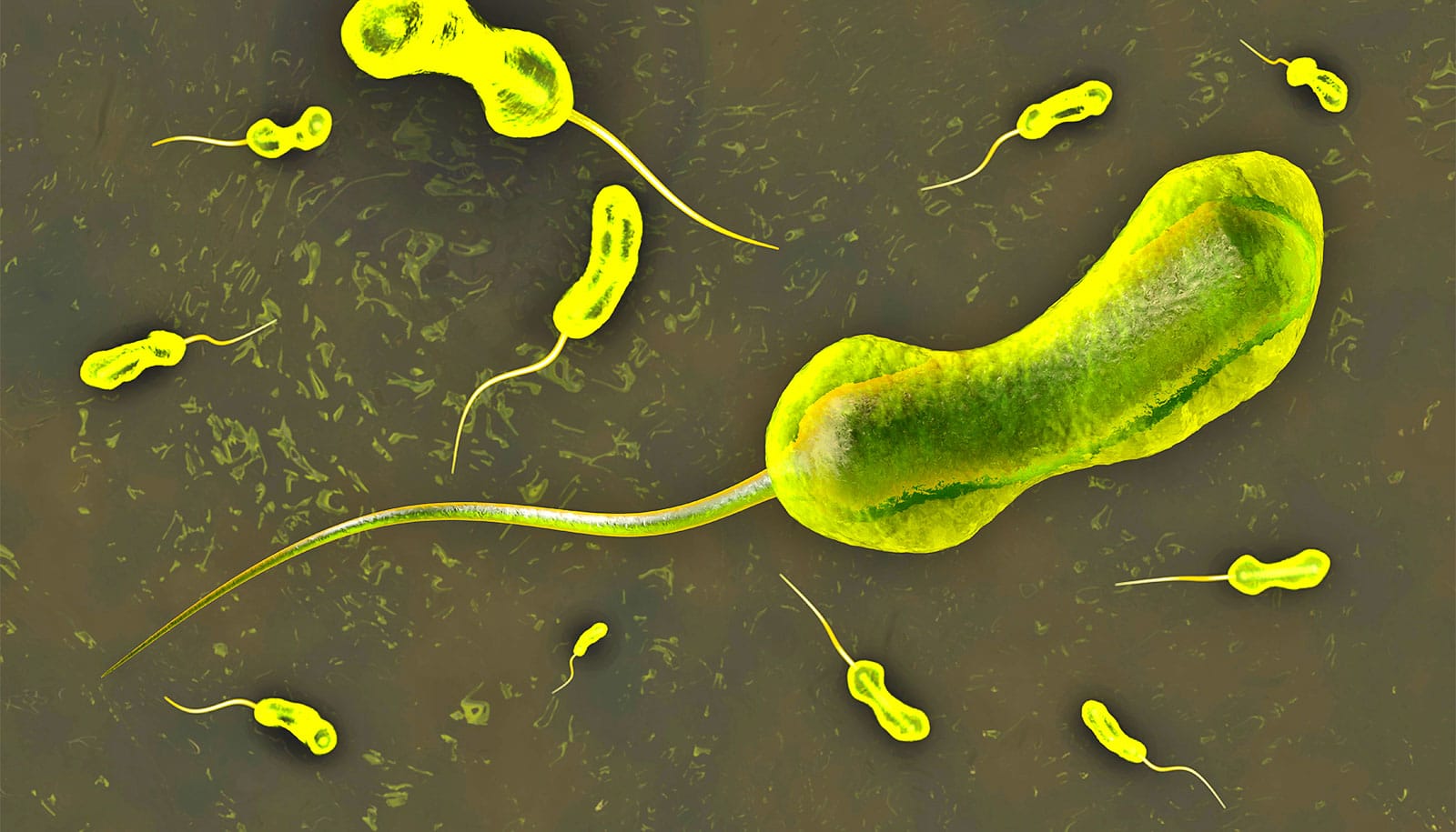
Vibrio cholerae, the bacterium responsible for cholera, relies on its flagellum for motility. This whip-like appendage allows the bacteria to navigate the watery environment of the small intestine and effectively reach and infect intestinal cells. The flagellum isn’t just about movement; its speed and force are crucial. Scientists believe this rapid motility helps V. cholerae penetrate the protective mucus layer lining the intestine, a key step in establishing infection. In fact, some cholera vaccines function by reducing bacterial motility, highlighting the flagellum’s importance in the infection process.
For decades, scientists have known the proteins that *make up* the flagellum, but visualizing its intricate structure – especially in its natural, functioning state – has been a major challenge. As Dr. Jun Liu, a professor of microbial pathogenesis at YSM and senior author of the study, explains, “To really understand the mechanism of the flagella—how they are able to assemble, how they rotate—you need near-atomic resolution.”
A 70-Year Mystery Unraveled
The unique structure of V. cholerae’s flagellum has long presented a hurdle to researchers. Unlike many othre bacteria, the flagellar proteins of V. cholerae are encased in a hydrophilic (water-loving) sheath. This sheath,while essential for the flagellum’s function,effectively shields the internal structure from traditional microscopic observation. Moreover, conventional methods for studying proteins frequently enough require killing the bacteria and breaking down its components, losing crucial details about how the flagellum operates within a living cell.
“The structure of V. cholerae’s flagella is a ‘70-year mystery,’” states Dr. Wangbiao Guo, a postdoctoral researcher in Liu’s lab and the study’s first author.To overcome these obstacles, the Yale team developed a novel microscopy technique. This involved genetically modifying V. cholerae to produce flagella proteins that would “light up” under specific conditions. The bacteria were then rapidly frozen in liquid ethane and examined using a powerful electron microscope, allowing for near-atomic-level visualization of the flagellum in its native state.
What the New Images Reveal
The resulting images revealed a surprising level of detail. while the core structure of the V. cholerae flagellum is similar to those found in other bacteria, the surface features are distinctly different. This suggests that V. cholerae’s flagellum has evolved unique adaptations within its protective sheath, contributing to its extraordinary speed and efficiency.
The research indicates that the flagellum rotates independently from the outer sheath. The hydrophilic nature of the sheath may create a lubricating layer, reducing friction and allowing for faster, more streamlined movement through the intestinal environment. This lubrication could be a key factor in V. cholerae’s ability to overcome the body’s natural defenses.
Implications for Future Treatments and Research
This breakthrough isn’t just about satisfying scientific curiosity; it has significant implications for the development of new cholera treatments. Understanding the precise structure and mechanics of the flagellum opens up possibilities for:
- Targeted Drug Development: Drugs could be designed to disrupt flagellar assembly or function,effectively immobilizing the bacteria and preventing infection.
- Improved Vaccine Strategies: A deeper understanding of flagellar structure could lead to the development of more effective vaccines that target this critical component of the bacteria.
- Novel Antimicrobial Approaches: The unique adaptations of the V. cholerae flagellum may present vulnerabilities that can be exploited by new antimicrobial agents.
dr. Liu emphasizes that this research is just the beginning. “We have at least provided some clues for the next development,” he says. Further research will focus on unraveling the precise mechanisms of flagellar rotation and the role of the sheath in facilitating movement. The techniques developed in this study will also be invaluable for investigating the flagella of other bacterial pathogens.
Cholera: A Global Health Challenge
Cholera is an acute diarrheal infection caused by the ingestion of food or water contaminated with V. cholerae. The disease can cause severe dehydration and, if left untreated, can be fatal within hours. while preventable and treatable, cholera remains a major public health concern in many parts of the world, particularly in areas with inadequate sanitation and limited access to clean water. According to the World Health Institution,countries in Africa and Asia bear the heaviest burden of the disease.
This new research offers a beacon of hope in the fight against cholera, providing a crucial piece of the puzzle in understanding how this deadly bacterium infects its host. By unlocking the secrets of the flagellum, scientists are one step closer to developing more effective strategies to combat this persistent global health threat.
Source: Yale