
Muscle Mass & Visceral fat Linked to Younger Brain age, Study Finds
New research suggests a strong connection between body composition – specifically muscle mass and visceral fat levels - and brain health, potentially impacting the risk of age-related cognitive decline. The findings, to be presented next week at the Radiological Society of North America (RSNA) annual meeting, indicate that individuals with greater muscle mass and a lower ratio of visceral fat to muscle mass tend to exhibit a younger “brain age.”
Visceral fat, the fat stored deep within the abdominal cavity surrounding internal organs, appears to be a key factor. Cyrus Raji, associate professor of radiology and neurology at Washington university School of Medicine in St. Louis, Missouri, and the study’s senior author, believes this link could translate to a reduced risk of neurodegenerative diseases like Alzheimer’s.
“Although we know chronological aging is associated with muscle loss and increased abdominal fat, this study demonstrates that these factors are directly related to brain aging itself,” Raji explained. ”Muscle mass and fat mass, as measured in the body, are key indicators of brain health and accompany the brain’s aging process.”
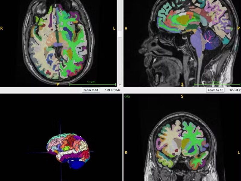
Researchers determined “brain age” using structural MRI scans, computationally estimating chronological age based on brain structure. Muscle mass, assessed via whole-body MRI, can serve as an indicator of interventions aimed at improving frailty and brain health. Brain age prediction from MRI scans can also provide insights into Alzheimer’s risk factors, such as muscle loss.
The study involved 1,164 healthy participants (52% women) from four different research sites. Participants had an average age of 55.17 years and underwent whole-body MRI scans utilizing T1-weighted sequences, a technique that clearly differentiates between fat and fluid, allowing for precise imaging of muscle, adipose tissue, and the brain.
An artificial intelligence (AI) algorithm was employed to quantify normalized total muscle volume, visceral fat, subcutaneous fat (fat under the skin), and ultimately, brain age. The analysis revealed a significant association: a higher ratio of visceral fat to muscle mass correlated with an older brain age. Interestingly, subcutaneous fat did not show a significant link to brain age.
Professor Raji emphasizes that both gaining muscle mass and reducing visceral fat are achievable goals. He believes whole-body MRI and AI-driven brain age estimates offer objective tools for monitoring the effectiveness of interventions designed to reduce visceral fat and preserve muscle mass.
The research also has implications for future therapies. Raji suggests the findings support the inclusion of body composition biomarkers in clinical trials evaluating metabolic interventions. He specifically points to the potential of GLP-1-based drugs, currently used for weight loss, but notes a need for careful consideration of their impact on muscle mass. Drugs like Ozempic, while effective for fat loss, “may also be related to a greater loss of muscle mass,” he cautioned.
Ultimately, Raji hopes this study will guide future research focused on quantifying body fat, muscle mass, and brain age via MRI, leading to optimized dosing regimens for GLP-1s and other treatments to maximize benefits for both body and brain health.