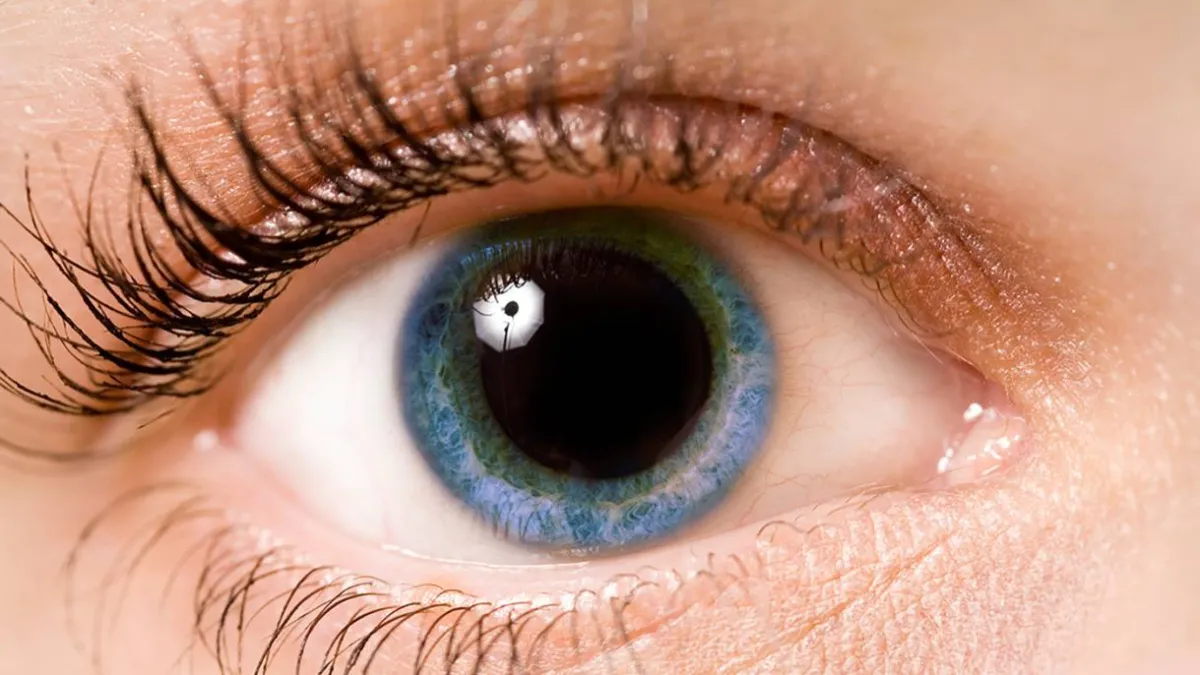
Why Do Pupils Dilate When Surprised
When an individual encounters sudden surprise, the human pupil dilates rapidly, a physiological reaction driven by the sympathetic nervous system. This involuntary biological shift forms part of the classic fight-or-flight response, preparing the body to process environmental threats or unexpected stimuli with heightened visual acuity.
Key Clinical Takeaways:
- Pupillary dilation during surprise is controlled by the sympathetic nervous system and the contraction of the dilator pupillae muscle.
- The response maximizes retinal light capture, enhancing an individual’s immediate visual field during acute stress.
- Understanding autonomic nervous system responses assists clinicians in evaluating neurological function and physiological stress markers.
Autonomic Regulation and the Dilator Pupillae Muscle
The mechanics of pupillary dilation, known clinically as mydriasis, rely on an intricate balance between two opposing smooth muscles within the iris. According to physiological research published by the National Institutes of Health (NIH), the sudden onset of an unexpected event triggers an immediate surge of adrenaline. This sympathetic activation stimulates the dilator pupillae muscle, causing the pupil to widen within milliseconds.
Simultaneously, parasympathetic signals from the Edinger-Westphal nucleus that normally constrict the pupil are temporarily inhibited. This dual-pathway shift ensures that the eye adapts instantly to changing environmental contexts. Patients or individuals noticing persistent abnormalities in pupillary response—such as unequal pupils or delayed reactivity—should schedule an evaluation with a qualified board-certified neurologist or ophthalmologist to rule out underlying cranial nerve pathologies.
Evolutionary Function of Rapid Visual Acquisition
From an evolutionary standpoint, widening the pupil during moments of surprise serves a distinct survival function. By increasing the aperture of the eye, more ambient light reaches the retina, heightening peripheral awareness and motion detection. Research indexed via PubMed demonstrates that this reflex optimizes visual processing speed, allowing the brain to categorize sudden stimuli faster than standard resting states permit.
This autonomic adjustment rarely occurs in isolation. It frequently accompanies other sympathetic indicators, including elevated heart rate, increased myocardial contractility, and transient vasoconstriction. Healthcare providers analyzing autonomic nervous system disorders often utilize pupillometry to measure the integrity of these reflex arcs. For comprehensive diagnostic assessments, patients can consult specialists through a trusted diagnostic imaging and neurology center.
Clinical Significance in Neurological and Psychological Evaluations
While normal pupillary reactivity to surprise or emotional shifts indicates a healthy autonomic nervous system, blunted or hyper-reactive responses can signal clinical concerns. Neurologists assess pupillary reflexes during routine physical examinations to evaluate brainstem integrity. Disruptions in the sympathetic pathway can point to conditions like Horner’s syndrome, whereas fixed, dilated pupils may indicate pharmacological agents, trauma, or increased intracranial pressure.
Managing complex neurological conditions requires coordinated care between primary practitioners and subspecialists. Clinicians seeking to streamline patient referrals or establish standardized diagnostic pathways often coordinate with a specialized medical practice management network to ensure seamless clinical oversight.
*Disclaimer: The information provided in this article is for educational and scientific communication purposes only and does not constitute medical advice. Always consult with a qualified healthcare provider regarding any medical condition, diagnosis, or treatment plan.*