Unicornuate Uterus With Rudimentary Horn Adenomyoma and Renal Agenesis: A Case Report
A rare congenital anomaly involving a unicornuate uterus with a rudimentary horn adenomyosis, ipsilateral renal agenesis, and deep infiltrating endometriosis presents significant diagnostic challenges in modern gynecology, according to a case report published in Cureus. Managing complex mullerian duct anomalies requires precise imaging modalities and personalized surgical planning to mitigate long-term patient morbidity and preserve reproductive health.
Key Clinical Takeaways:
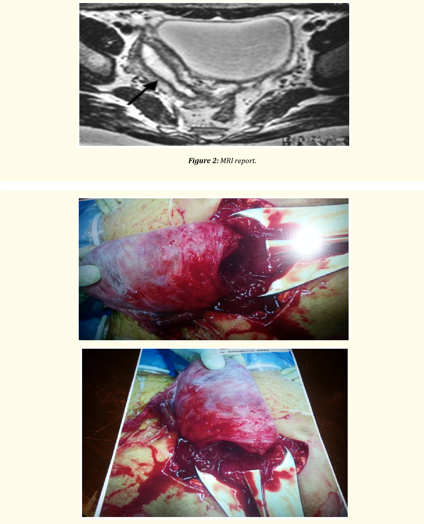
- The patient presented with a rare triad of a unicornuate uterus, rudimentary horn adenomyosis, and ipsilateral renal agenesis, complicated by deep infiltrating endometriosis.
- Accurate diagnosis relied on advanced cross-sectional imaging and diagnostic laparoscopy to map abnormal pelvic anatomy.
- Surgical excision of the rudimentary horn and thorough management of deep infiltrating endometriosis form the current standard of care for symptomatic presentations.
Understanding Mullerian Duct Anomalies and Associated Pathogenesis
Mullerian duct anomalies encompass a spectrum of congenital malformations resulting from abnormal development, fusion, or resorption of the paramesonephric ducts during embryogenesis. A unicornuate uterus represents the failure of one duct to develop fully, frequently co-occurring with unilateral renal anomalies due to the close embryological relationship between the genital and urinary systems. The presence of a rudimentary horn—whether communicating or non-communicating with the main uterine cavity—introduces severe clinical risks, including dysmenorrhea, chronic pelvic pain, and rudimentary horn pregnancy rupture. When complicated by deep infiltrating endometriosis and adenomyosis within the rudimentary structure, the pathogenesis involves complex hormonal and anatomical interactions that exacerbate patient suffering.
According to findings documented in PubMed Central, recognizing these overlapping pathologies early prevents misdiagnosis and reduces the risk of acute surgical emergencies. Clinicians evaluating patients with persistent unilateral pelvic pain and congenital renal absence must maintain a high index of suspicion for associated Mullerian malformations. Comprehensive diagnostic workups typically involve pelvic magnetic resonance imaging and transvaginal ultrasonography to delineate uterine architecture and identify functional endometrial tissue within non-communicating horns.
Diagnostic Pathways and Surgical Management Strategies
Mapping complex pelvic anatomy demands a multidisciplinary approach involving gynecologic surgeons, diagnostic radiologists, and fertility specialists. When deep infiltrating endometriosis involves retroperitoneal structures or the bowel, coordinating care through specialized centers becomes essential for optimal outcomes. Patients navigating these intricate diagnostic phases can benefit greatly from expert evaluation by connecting directly with a board-certified gynecologic laparoscopic surgeon to review advanced imaging and discuss individualized surgical interventions.
Surgical excision of the rudimentary horn remains the definitive treatment to eliminate the risk of ectopic pregnancy and alleviate severe dysmenorrhea caused by entrapped menstrual blood. Concurrently, meticulous excision of deep infiltrating endometriosis lesions restores normal pelvic architecture and relieves chronic inflammatory symptoms. Healthcare providers managing complex surgical candidates often collaborate with multidisciplinary pelvic pain and endometriosis clinics to ensure comprehensive postoperative recovery and long-term surveillance.
Future Directions in Congenital Pelvic Pathology Research
Advancements in three-dimensional imaging and genetic screening continue to refine the early detection of complex reproductive tract anomalies. As clinical registries expand, researchers aim to better understand the molecular links between congenital uterine malformations and the development of severe phenotypes like deep infiltrating endometriosis. Continued investment in specialized diagnostic networks ensures that patients with multifaceted anatomical presentations receive timely, targeted interventions.
For individuals seeking specialized diagnostic evaluations or personalized treatment planning for congenital pelvic anomalies, scheduling a consultation with a specialized reproductive pelvic surgery center provides access to expert multidisciplinary care.
*Disclaimer: The information provided in this article is for educational and scientific communication purposes only and does not constitute medical advice. Always consult with a qualified healthcare provider regarding any medical condition, diagnosis, or treatment plan.*