Understanding the Clinical Process of Skin Cancer Diagnosis
When facing a skin cancer diagnosis, patients frequently encounter a stark gap between an eleven-minute clinical consultation and the complex medical decisions that follow in the parking lot. Knowing precisely what questions to ask a dermatologist or oncologist turns a brief appointment into a structured conversation, ensuring final treatment choices are grounded in a clear understanding of pathology reports, recovery timelines.
- Patients should routinely request a physical copy of their pathology report and ask their dermatologist to walk through five core items: cancer type, subtype, tumor depth (such as Breslow thickness for melanoma), margin status, and mitotic rate.
- Under NHS guidance cited in the source materials, the two-week rule mandates that general practitioners make urgent suspected-cancer referrals for melanoma or squamous cell carcinoma so patients see a specialist within 14 days.
- Depending on the lesion’s severity and location, care teams may involve dermatologists, Mohs micrographic surgeons, surgical or medical oncologists, and radiation oncologists in a multidisciplinary review.
Dermatoscopes and Biopsies Lead to Pathology Reports
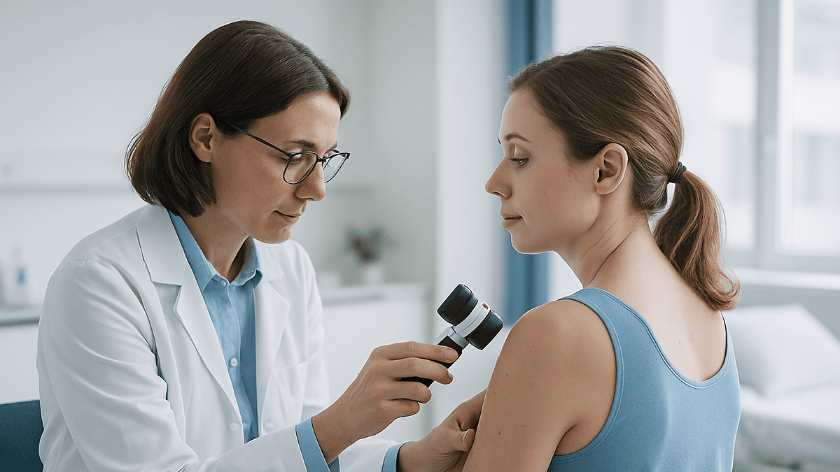
The clinical route from a suspicious spot to an established treatment plan begins with a physical examination via dermatoscope, a handheld polarized magnifier that exposes subsurface pigment patterns. According to the foundational workflow detailed in directory-sourced guidance, if a lesion looks clinically suspicious, the definitive next step is a biopsy where a local anesthetic is applied to remove all or part of the tissue for microscopic evaluation by a pathologist. Moffitt Cancer Center notes that patients should immediately ask their specialist what specific type of skin cancer they have, whether it has spread, and what stage the disease has reached. The resulting written pathology report forms the absolute hinge of all subsequent medical management. It identifies the exact cancer type—such as basal cell carcinoma, which grows slowly and rarely metastasizes, versus squamous cell carcinoma or more aggressive melanoma—and describes the Breslow thickness, measuring the precise distance in millimeters from the skin surface to the deepest invading malignant cell.
Timeframes for specialized evaluation vary significantly depending on institutional protocols and clinical urgency. The United Kingdom operates under a strict two-week rule where general practitioners submit urgent suspected-cancer referrals for patients presenting with clinical signs of melanoma or squamous cell carcinoma, ensuring specialist evaluation within 14 days to prevent delays that impact lesion depth and tumor staging. Conversely, suspected basal cell carcinomas are typically routed through routine non-urgent pathways due to their slow growth kinetics. Patients managing changing or persistent lesions are advised to verify whether their referral is marked urgent, maintain photographic documentation of the spot to track morphological evolution over time, or inquire if teledermatology remote assessments can accelerate the triage process.
Assembling the Multidisciplinary Care Team
While standard basal cell and minor squamous cell carcinomas are routinely diagnosed and excised directly in a dermatologist’s office, advanced presentations require a broader clinical cast. Mohs micrographic surgeons perform specialized layered excisions with immediate microscopic margin checks for complex facial tumors. When a tumor invades regional lymph nodes or requires systemic management, surgical oncologists, medical oncologists specializing in immunotherapy or targeted drug regimens, and radiation oncologists join the treating team. Moffitt Cancer Center advises patients to ask whether their case will be evaluated through a multidisciplinary review, how many times their specific surgeon has performed the proposed procedure, and whether a second opinion should be sought before initiating intervention.
Treatment Selection Depends on Tumor Morphology and Location
Therapeutic selection depends entirely on tumor morphology, anatomical location, and patient comorbidities. Standard surgical excisions, specialized micrographic surgery, topical prescription creams, radiation therapy, and systemic immunotherapies each carry distinct recovery windows, scar profiles, and side-effect management protocols. Before committing to a pathway, patients must establish clear recovery expectations and discuss recurrence monitoring schedules. As outlined in directory source material, follow-up parameters dictate how frequently routine skin cancer screenings must be scheduled in the future, providing a structured safety net long after initial surgical healing is complete.
Disclaimer: The information provided in this article is for educational and scientific communication purposes only and does not constitute medical advice. Always consult with a qualified healthcare provider regarding any medical condition, diagnosis, or treatment plan.