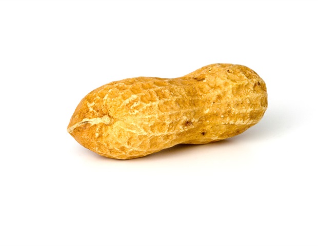
UNC Researchers Link Missing Gut Protein to Peanut Allergy Susceptibility
Researchers at the University of North Carolina School of Medicine have identified a specific cellular defect in the gut lining that may predispose children to peanut allergies before they are ever exposed to the allergen. The findings, published September 30 in Cellular and Molecular Gastroenterology and Hepatology, suggest that a lack of the enzyme lysozyme 1 (Lyz1) in specialized gut cells creates a biological environment conducive to allergic sensitization.
- Paneth cells, which act as antimicrobial sentinels in the gut, show a critical deficiency in the enzyme lysozyme 1 in allergy-susceptible models.
- This genetic defect alters the gut microbiome and immune surveillance, priming the intestinal barrier for allergic reactions before initial peanut exposure.
- Human biopsies confirm that pediatric patients with peanut allergies exhibit a significant reduction in lysozyme-positive crypts compared to non-allergic individuals.
Genetic Origins of Paneth Cell Dysfunction
The study, led by graduate student Katelyn Clough and Dr. Shehzad Z. Sheikh and Dr. Erin C. Steinbach, utilized single-cell RNA sequencing to map the intestinal lining of allergy-susceptible CC027 mice. The researchers discovered that these mice possess a genetic variant inherited from the wild mouse species CAST/EiJ that lacks the Lyz1 gene entirely. This results in the complete absence of lysozyme 1 production in Paneth cells.
According to the UNC School of Medicine, this is not a phenomenon isolated to murine models. When the research team analyzed small intestinal biopsies from pediatric patients, they observed a statistically significant decrease in the number of lysozyme-positive crypts in those with peanut allergies. This indicates that the loss of this antimicrobial protein is a biologically relevant feature of human susceptibility to peanut allergies.
Epithelial Remodeling Impairs Gut Immune Surveillance
Beyond the absence of lysozyme, the researchers identified a broader pattern of epithelial remodeling that occurs in the absence of allergen exposure. The atlas revealed a depletion of interferon-responsive absorptive enterocytes (AE-IFN), suggesting that the gut’s antiviral and immune surveillance programs are impaired in allergy-susceptible subjects. The study also noted an expansion of goblet cells—which produce protective mucus—and a reduction in enteroendocrine cells, which regulate gut permeability.
Tuft cells, known to initiate type 2 allergic immune responses, were found to be increased in number and showed higher expression of allergic immune receptors. These findings suggest that the intestinal epithelium is not merely a passive barrier but an active driver of allergic pathogenesis. The study, supported by the Center for Gastrointestinal Biology and Disease and the Division of Rheumatology, Allergy & Immunology, highlights a mechanistic chain where a single missing protein triggers a cascade of microbiome shifts and immune skewing.
Lysozyme Deficiency Alters Gut Microbiome Composition
The deficiency of lysozyme 1 has direct consequences for the composition of the gut microbiome. The research indicates that the lack of this enzyme leads to an enrichment of bacterial genera such as Ruminococcus and Akkermansia. This altered microbial profile is associated with increased intestinal permeability and a heightened state of allergic immune skewing.
Current management for the approximately 1.6 million children in the United States affected by peanut allergy remains limited to strict avoidance and the use of emergency epinephrine. Identifying these gut barrier defects early may provide a new window for clinical intervention.
For families managing life-threatening food allergies, the research shows how complex the condition is. Clinical experts emphasize that the next phase of this research will involve larger pediatric cohorts and the use of human intestinal organoid models. These models will test whether restoring lysozyme levels or correcting the gut microbiome can effectively repair barrier function.