Tuberculous Posterior Mediastinal Neuroenteric Cyst with Secondary Autoimmune Hemolytic Anemia: A Case Report
A recent medical case report published in the open-access journal Cureus details an unusual clinical presentation where a tuberculous infection within a posterior mediastinal neuroenteric cyst triggered secondary autoimmune hemolytic anemia (AIHA). The patient presented diagnostic and therapeutic challenges, highlighting rare extrapulmonary manifestations of tuberculosis coupled with autoimmune hematologic complications.
Key Clinical Takeaways:
- A rare case of a posterior mediastinal neuroenteric cyst became infected with tuberculosis, leading to secondary autoimmune hemolytic anemia.
- The patient’s complex presentation required coordinated evaluation by thoracic surgery, infectious disease specialists, and hematologists.
- Management centered on targeted antimicrobial therapy combined with surgical intervention to address the infected mediastinal mass.
Clinical Presentation and Diagnostic Challenges
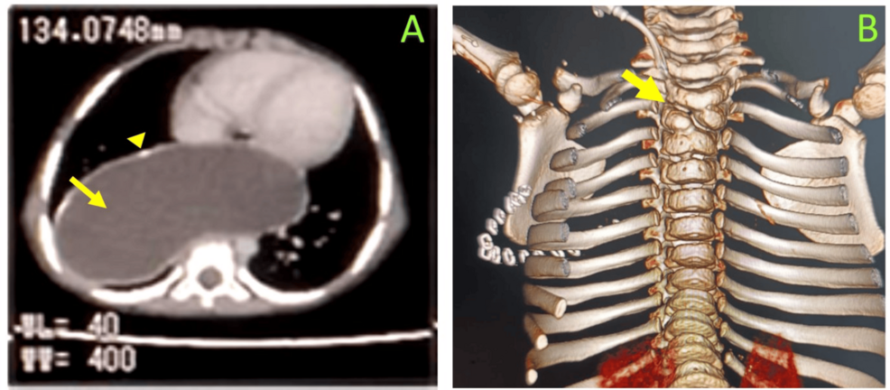
Neuroenteric cysts are rare developmental congenital anomalies resulting from abnormal separation of the notochord from the endoderm, typically presenting as posterior mediastinal masses. While these cysts are usually benign and often asymptomatic until they cause local compression, infection within the cyst cavity introduces severe clinical complications. According to the case report published in PubMed indexed literature, the nidus of infection provided by the tuberculous process initiated an aberrant immune response. This systemic inflammatory activation resulted in secondary autoimmune hemolytic anemia, a condition where the immune system mistakenly targets and destroys red blood cells.
Diagnosing concurrent mediastinal tuberculosis and secondary AIHA demands rigorous diagnostic investigation. Standard imaging modalities, including contrast-enhanced computed tomography, help delineate the anatomical boundaries of the posterior mediastinal cyst and assess airway or vascular compression. However, identifying the underlying infectious etiology often requires tissue biopsy or surgical excision followed by mycobacterial cultures and polymerase chain reaction assays. When patients present with unexplained hemolytic anemia alongside a cystic mediastinal lesion, clinicians must maintain a high index of suspicion for atypical or infectious etiologies driving the autoimmune response.
Therapeutic Interventions and Multidisciplinary Management
Managing dual pathologies involving an active infectious source and an immune-mediated hematologic disorder requires a synchronized therapeutic approach. Standard protocols for extrapulmonary tuberculosis rely on multi-drug anti-tubercular regimens to eradicate the mycobacterial load. Concurrently, managing secondary AIHA often involves corticosteroids or other immunomodulatory agents to suppress red blood cell destruction while the primary trigger is treated.
Surgical resection remains the definitive treatment for symptomatic or infected neuroenteric cysts to prevent recurrent infection, mediastinitis, or compressive symptoms. Post-operative recovery and long-term stabilization depend heavily on specialized monitoring. For clinicians and medical facilities managing complex thoracic anomalies, consulting with vetted specialists is essential. Patients and referring physicians can coordinate comprehensive evaluations by connecting with a board-certified thoracic surgeon or an infectious disease specialist to ensure optimal therapeutic drug monitoring and surgical follow-up.
Implications for Future Clinical Practice
This documented intersection of a rare congenital cyst, mycobacterial infection, and autoimmune hematologic phenomena underscores the diagnostic complexity frequently encountered in modern clinical practice. Clinicians evaluating atypical mediastinal masses must consider not only local mass effects but also systemic paraneoplastic or immunological manifestations. As medical literature continues to document these atypical presentations, healthcare systems rely on stringent diagnostic accuracy and multidisciplinary coordination.
Navigating complex diagnostic workups and ensuring adherence to established clinical guidelines requires robust institutional support. Healthcare providers seeking guidance on regulatory compliance, clinical trial integration, or specialized diagnostic pathways often collaborate with healthcare compliance consultants and advanced diagnostic pathology laboratories to streamline patient care pathways and mitigate diagnostic delays.
*Disclaimer: The information provided in this article is for educational and scientific communication purposes only and does not constitute medical advice. Always consult with a qualified healthcare provider regarding any medical condition, diagnosis, or treatment plan.*