Surgeons Successfully Repair Massive 13.5-cm Atherosclerotic Aortic Aneurysm
medical literature documented a rare surgical intervention for a 70-year-old woman presenting with a 13.5-centimeter giant atherosclerotic ascending aortic aneurysm with aortic arch involvement. The patient, who exhibited progressive severe dyspnea, generalized fatigue, and hemodynamic compromise, underwent emergency open surgical repair involving supracoronary ascending aortic replacement and total arch replacement under deep hypothermic circulatory arrest.
Key Clinical Takeaways:
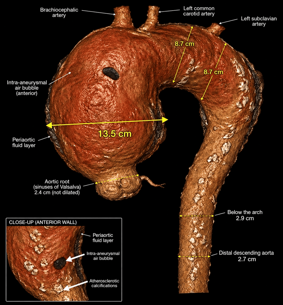
- A 70-year-old female patient presented with a giant ascending aortic aneurysm measuring 13.5 cm, nearly five times the normal diameter, alongside an 8.7 cm aortic arch aneurysm.
- Emergency open surgical repair successfully utilized valve-sparing total arch replacement, deep hypothermic circulatory arrest, and separate supra-aortic branch reconstruction with a branched Dacron graft.
- Histopathology confirmed an atheromatous aneurysm with thrombotic degeneration, while microbiological studies ruled out infection, highlighting an exceptionally rare presentation of atherosclerosis in the ascending aorta.
Patient Arrives with Massive Ascending Aortic Aneurysm
International guidelines typically recommend surgical intervention for ascending aortic aneurysms at diameters reaching or exceeding 5.5 cm due to sharply escalating risks of rupture, dissection, and mortality. The patient’s aneurysm measuring 13.5 cm in the ascending aorta and 8.7 cm in the aortic arch pushed far beyond standard operative thresholds. reported that the patient arrived at the emergency department experiencing New York Heart Association Class IV dyspnea, hypotension, and respiratory distress, accompanied by a C-reactive protein level of 231.3 mg/L and a hemoglobin count of 10.5 g/dL.
Diagnostic Imaging and Pathological Findings
Contrast-enhanced chest computed tomography scans detailed the massive dilation extending through the ascending aorta and into the arch, while sparing the aortic root and descending thoracic aorta. Periaortic fluid and specific imaging features indicated an impending rupture. While thoracic aortic aneurysms frequently stem from medial degeneration, connective tissue disorders, or bicuspid aortic valve disease, histopathological examination of this specific case confirmed an atheromatous aneurysm characterized by thrombotic degeneration. Microbiological testing subsequently excluded any infectious etiology.
Surgical Team Performs Total Arch Replacement
Managing disease of this scale demanded advanced, anatomy-driven surgical techniques. The surgical team performed supracoronary ascending aortic replacement combined with a total arch replacement. This approach relied on deep hypothermic circulatory arrest to protect cerebral and visceral organs during the cessation of blood flow. Separate reconstruction of the supra-aortic branches was achieved utilizing a branched Dacron graft. This technique successfully accomplished durable exclusion of the extensive pathology while preserving the patient’s native aortic root structures.
Disclaimer: The information provided in this article is for educational and scientific communication purposes only and does not constitute medical advice. Always consult with a qualified healthcare provider regarding any medical condition, diagnosis, or treatment plan.