
New ‘ExoPatch‘ Detects Melanoma Through skin, Offers Potential for Early Cancer Diagnosis
ANN ARBOR, MI – Researchers at the university of Michigan have developed a novel patch, dubbed the ExoPatch, capable of detecting melanoma through the skin by capturing and analyzing exosomes – tiny vesicles released by cells – present in the fluid beneath the skin’s surface. The technology, detailed in a recent study published in Biosensors and Bioelectronics, demonstrates a potential pathway for earlier and less invasive cancer diagnosis.
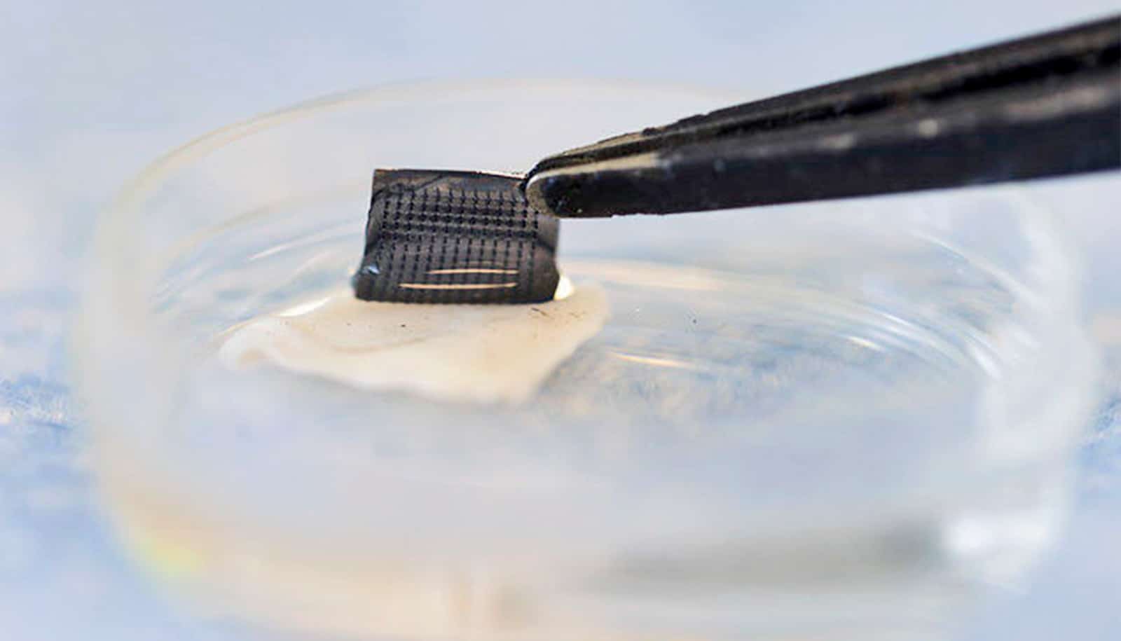
The ExoPatch utilizes an array of microneedles, each approximately 600 nanometers in length, to gently access fluid from the interstitial space – the area between skin cells. For context, the epidermis of the human forearm is roughly 18,300 nanometers thick. The patch is coated with a gel designed to capture exosomes released by both healthy and cancerous cells.
To validate the ExoPatch’s ability to isolate melanoma exosomes,the research team conducted experiments using mouse skin samples. Half of the samples were from healthy mice, while the other half came from mice injected with a fragment of a human melanoma tumor. Following a 15-minute request of the ExoPatch, microscopic analysis revealed that exosomes adhered effectively to the microneedles, falling within the expected size range of 30 to 150 nanometers.
“When looking at microscopy images,I was happy to see how nicely the exosomes adhered to the microneedles and were within the 30 to 150 nanometer size range we expect,” said Scott Smith,a doctoral student of chemical engineering and co-lead author of the study.
After exosome capture, the gel was dissolved, and the resulting sample was analyzed using test strips. These strips successfully differentiated between melanoma and healthy tissue, exhibiting a 3.5-fold darker line in samples originating from melanoma tissue. Furthermore, the ExoPatch demonstrated a important ability to specifically target cancerous exosomes, isolating 11.5 times more exosomal protein from melanoma tissue compared to healthy tissue.
The research team, led by Sunita Nagrath, a professor of chemical engineering, plans to initiate a pilot study in humans, followed by thorough clinical trials, to pave the way for widespread clinical application. Nagrath’s lab focuses on developing liquid biopsy technologies for early cancer detection.
Beyond melanoma, the ExoPatch’s gel coating can be adapted to detect exosomes released by other cancers characterized by solid tumors, including lung, breast, colon, prostate, and brain cancer (specifically glioblastoma).
“This is the first patch designed to capture disease-specific exosomes from fluid under the skin. The potential applications are huge,” Nagrath stated.
The research was funded by the National Institutes of Health. The device fabrication occurred at the Lurie Nanofabrication Facility, with testing partially conducted in the Biomedical Research Core Facilities and Rogel Cancer Center Immunology Core at the University of Michigan. The team has filed for patent protection with support from U-M Innovation Partnerships.