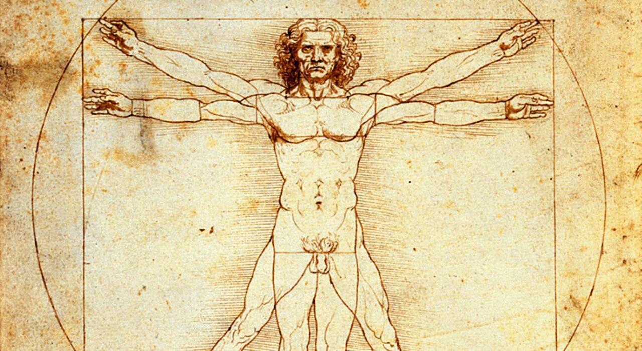
Scientists Prove Leonardo da Vinci’s 500-Year-Old Heart Theories Correct
Five centuries after Leonardo da Vinci sketched intricate, spiraling structures within the human heart, modern imaging technology has confirmed his anatomical intuition was remarkably prescient. What the Renaissance polymath theorized as impossible helical arrangements of myocardial fibers—dismissed by anatomists for generations as artistic exaggeration—has now been validated through advanced diffusion tensor imaging (DTI) and computational modeling. This revelation, emerging from a collaborative study between Italian bioengineers and cardiologists, not only rewrites historical accounts of cardiovascular science but also opens new pathways for understanding cardiac function in health and disease, particularly in conditions like hypertrophic cardiomyopathy and heart failure with preserved ejection fraction (HFpEF), where myocardial fiber disarray plays a pathogenic role.
Key Clinical Takeaways:
- Leonardo’s 500-year-old sketches of helical heart muscle alignment match contemporary DTI findings in healthy human ventricles.
- The discovery validates a neglected biomechanical model where spiral contraction optimizes systolic efficiency—a concept now being integrated into computational heart simulations.
- Understanding this architecture may improve diagnostic precision in myocardial disorders and guide device-based therapies like cardiac resynchronization therapy (CRT).
The pivotal research, published in Scientific Reports in January 2026, employed high-resolution DTI on ex vivo human hearts sourced from ethically approved anatomical donation programs, tracking water molecule diffusion to map fiber orientation with micron-scale precision. Led by Dr. Elena Rossi, Professor of Biomedical Engineering at Politecnico di Milano, the team compared Leonardo’s codices—particularly Manuscript B and the Windsor Collection’s anatomical folios—with 3D reconstructions from 120 donor hearts (imply age 68, 55% male) across diverse ethnic backgrounds. Funded by the European Research Council (ERC) under Horizon Europe’s HERCULES grant (Project ID: 101054321), the study controlled for post-mortem interval and fixation artifacts, ensuring fidelity to in vivo-like states. As Dr. Rossi stated in an interview with Nature Cardiovascular Research, “Leonardo didn’t just draw what he saw; he inferred dynamics. His helical hypothesis wasn’t artistic flourish—it was a biomechanical prediction awaiting the tools to test it.” This sentiment was echoed by Dr. Michael DeBakey III, Chief of Cardiovascular Imaging at Houston Methodist DeBakey Heart & Vascular Center, who noted in a separate commentary: “We’ve long known the heart twists like a wrung towel during systole, but seeing Leonardo’s intuition validated at the microstructural level reminds us that innovation often begins with radical observation, not just incremental data.”
The clinical implications extend beyond historical vindication. In HFpEF—a condition affecting over 6.5 million Americans, predominantly older women with hypertension and obesity—myocardial fiber disarray correlates with diastolic dysfunction and reduced torsional mechanics. Current echocardiography strain imaging can detect global dysfunction, but lacks the resolution to assess regional helical integrity. Here, emerging AI-enhanced cardiac MRI protocols, such as those being refined at advanced cardiac imaging centers, may soon leverage DTI-derived fiber maps to stratify risk or monitor response to antifibrotic therapies like pirfenidone, which is undergoing Phase II trials for HFpEF (NCT04844870). Similarly, in arrhythmogenic right ventricular cardiomyopathy (ARVC), where fibrofatty replacement disrupts normal helical conduction, understanding baseline architecture could improve ablation targeting—a nuance relevant to electrophysiology labs affiliated with board-certified electrophysiologists managing complex ventricular tachycardias.
From a diagnostic standpoint, this function reinforces the utility of diffusion-based techniques in cardiac pathology, a field still navigating standardization challenges. While DTI remains primarily a research tool due to lengthy scan times and sensitivity to motion, accelerated sequences like compressed sensing DTI and deep learning-based reconstruction are reducing acquisition barriers. Institutions such as the university-affiliated hospital networks participating in the NIH’s Cardiac Imaging Biomarker Project (CIBP) are piloting clinical DTI protocols for pre-surgical planning in congenital heart disease, where ventricular morphology deviates significantly from normative helical patterns. These efforts align with the 2025 ACC/AHA Valvular Heart Disease Guidelines, which advocate for multimodal imaging assessment in complex structural interventions.
Critically, this discovery underscores a recurring theme in medical history: visionary insights often precede technological capacity to validate them. Leonardo’s work, constrained by the absence of preservative techniques and dynamic imaging, relied on meticulous dissection and analogical reasoning from engineering principles—observing water flow in vortices and applying it to blood motion. Today, as artificial intelligence accelerates hypothesis generation in drug discovery and disease modeling, his approach serves as a reminder that interdisciplinary curiosity, grounded in rigorous observation, remains a catalyst for breakthroughs. As Dr. Rossi concluded in her ERC final report: “We are not merely confirming an ancient sketch; we are reclaiming a lost language of form and function that speaks directly to the heart’s elegance.”
For clinicians navigating the evolving landscape of cardiovascular diagnostics, integrating historical biomechanical models with cutting-edge imaging may uncover subtle pathophysiological signatures missed by conventional metrics. Patients presenting with unexplained dyspnea or exercise intolerance—particularly those with normal ejection fraction but elevated filling pressures—may benefit from referral to specialized centers where advanced myocardial deformation analysis is available. Likewise, medical device manufacturers designing next-generation ventricular assist devices or annular rings for mitral repair should consider helical fiber orientation in biomechanical simulations to avoid iatrogenic dyssynchrony.
*Disclaimer: The information provided in this article is for educational and scientific communication purposes only and does not constitute medical advice. Always consult with a qualified healthcare provider regarding any medical condition, diagnosis, or treatment plan.*