
Researchers at Osaka City University Graduate School of Medicine have developed a method using tomosynthesis – a form of 3D X-ray imaging – to detect scapular notch defects following reverse total shoulder arthroplasty (rTSA). The technique, detailed in a presentation at the 12th CAOS Japan Annual Meeting, aims to improve post-operative assessment of shoulder replacements.
Reverse total shoulder arthroplasty is increasingly used to treat glenohumeral arthritis, particularly in patients with rotator cuff deficiency. A key challenge following rTSA is identifying and addressing glenoid bone defects, which can contribute to implant loosening and pain. Traditional X-ray imaging often provides limited visualization of the scapular notch, a critical area for assessing bone quality.
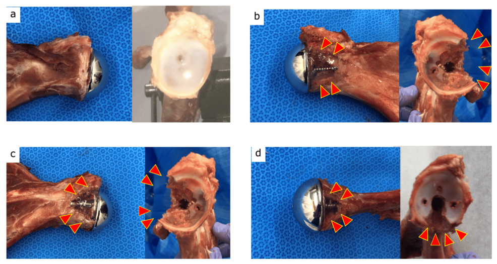
Yoshihiro Hirakawa, M.D., Ph.D., of Osaka City University, led the research, focusing on the application of tomosynthesis to improve the detection of these defects. Tomosynthesis generates cross-sectional images of the shoulder, offering a more detailed view of the glenoid compared to standard two-dimensional X-rays. The study utilized a pig model to simulate human shoulder anatomy and assess the accuracy of tomosynthesis in identifying scapular notch defects.
The development of improved imaging techniques for rTSA is an area of ongoing research. A 2017 systematic review published in the journal Joints identified a need for better research models, including biomechanical and virtual simulators, to study reverse total shoulder arthroplasty. The review, authored by Petrillo et al., examined existing literature using keywords such as “reverse total shoulder arthroplasty” and “research models.”
Recent studies have also focused on optimizing implant design to improve bone incorporation and reduce loosening. Research published in the Journal of Shoulder and Elbow Surgery in 2023 compared angled bony-increased offset implants with metal-augmented baseplate designs in 94 patients undergoing rTSA, following them for at least two years. The study assessed bone incorporation rates and implant loosening, identifying specific sites of potential failure.
Computational modeling is also being used to understand bone remodeling after rTSA. A study published on arXiv in 2017 applied a fully-nonlinear remodeling theory to investigate density changes following the procedure, providing new insights into the biological response to the implant. This research builds on previous perform by Kuhl and Steinmann on bone remodeling theory.
The Osaka City University research does not currently indicate when the tomosynthesis technique will be widely available for clinical use, nor does it detail the specific sensitivity and specificity of the method in detecting different sizes or types of scapular notch defects. Further research and clinical trials will be necessary to validate the technique and establish its role in routine post-operative care.