Posterior Reversible Encephalopathy Syndrome Mimicking Ischaemic Stroke in Severe Autoimmune Hyperthyroidism
A woman presenting with severe autoimmune thyrotoxicosis developed posterior reversible encephalopathy syndrome complicated by recurrent seizures and profound cortical visual loss, according to a case report. Medical records showed her initial occipital MRI abnormality was interpreted as a posterior cerebral artery territory infarction before follow-up imaging demonstrated substantial interval improvement characteristic of posterior reversible encephalopathy syndrome.
-
Key Clinical Takeaways:
- The patient presented with altered mental status, severe thyrotoxicosis, and fever, initially raising concerns for a thyrotoxic crisis or sepsis.
- Serial electroencephalograms confirmed focal-onset subclinical seizures and ongoing focal-onset seizure activity that resolved following antiepileptic escalation with levetiracetam, intravenous phenytoin, and lacosamide.
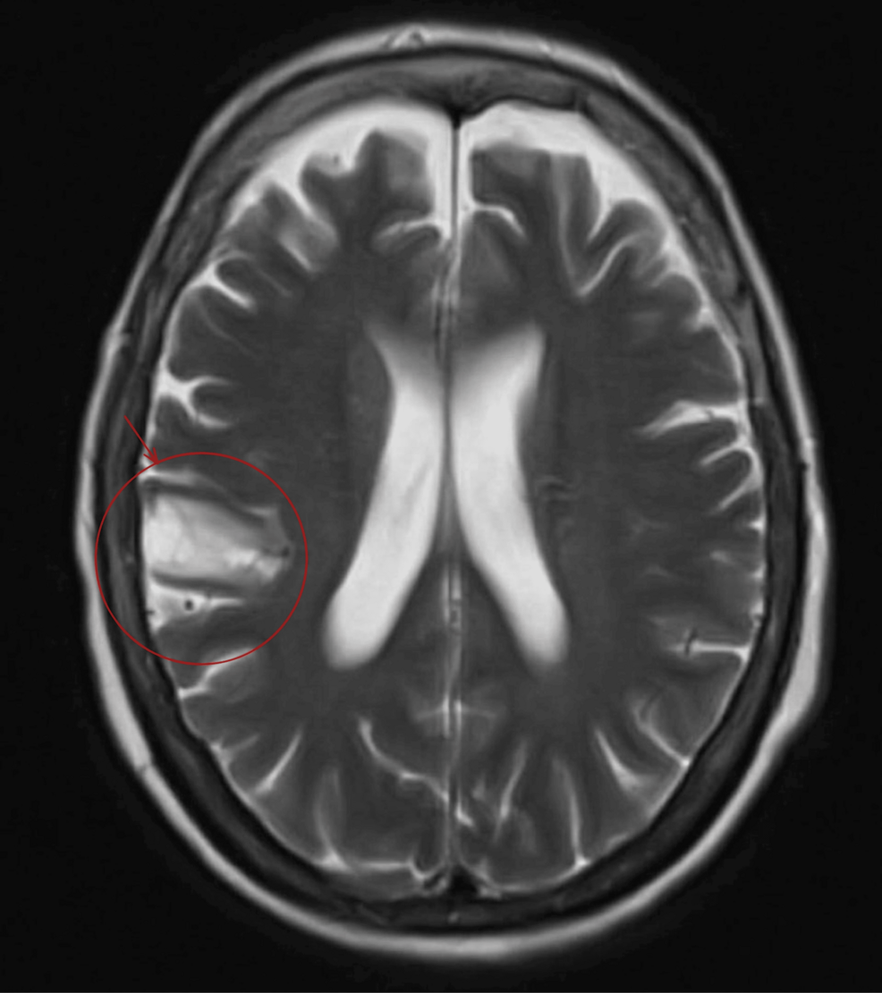
- Brain magnetic resonance imaging revealed cortical and subcortical T2/FLAIR hyperintensity in the right occipital lobe with diffusion restriction, mimicking an acute ischaemic stroke until interval imaging confirmed posterior reversible encephalopathy syndrome.
Patient Arrives Confused with Tachycardia and Fever
The patient was brought to the emergency department after being found collapsed on her bedroom floor. Upon arrival, her Glasgow Coma Scale score was 15 out of 15, though she appeared confused and responsive only to verbal stimuli. Initial vital signs demonstrated a sinus tachycardia of 118 beats per minute, an oxygen saturation of 90 percent on room air, a respiratory rate of 18 breaths per minute, and a blood pressure of 92 over 39 millimeters of mercury. She subsequently developed a fever reaching a peak temperature of 38.7 degrees Celsius.
Arterial blood gas analysis indicated a mixed respiratory acidosis with hypercapnia, showing a pH of 7.31 and a partial pressure of carbon dioxide of 7.16 kilopascals, alongside a normal lactate level of 1.17 millimoles per liter. Blood tests revealed an acute kidney injury with a creatinine of 168 micromoles per liter and an estimated glomerular filtration rate of 29 milliliters per minute per 1.73 square meters, alongside anaemia showing a haemoglobin level of 93 grams per liter. Thyroid function tests demonstrated severe thyrotoxicosis characterized by a suppressed thyroid-stimulating hormone level below 0.008 milli-international units per liter, a markedly elevated free thyroxine level of 34.5 picomoles per liter, and a free triiodothyronine level of 29.7 picomoles per liter. Strongly positive thyroid peroxidase and thyroid-stimulating hormone receptor antibodies confirmed a diagnosis of Graves’ disease.
Respiratory Failure Leads to Intensive Care Admission
Despite initial resuscitation with intravenous fluids and broad-spectrum antibiotics, the patient’s neurological and respiratory status deteriorated. Her Glasgow Coma Scale score dropped to 12 out of 15, accompanied by worsening oxygen requirements and an inability to maintain her airway independently. She developed severe hypercapnic type 2 respiratory failure with mixed respiratory and metabolic acidosis. A computed tomography pulmonary angiography ruled out a pulmonary embolism, while a head computed tomography scan demonstrated established low attenuation in the right parietal lobe and right basal ganglia consistent with previous infarcts, showing no acute haemorrhage or mass effect.
Following her transfer to the intensive care unit, non-invasive ventilation via bilevel positive airway pressure was initiated, which improved her respiratory acidosis and hypercapnia without the need for vasopressor support. Due to potential neck stiffness observed early in the admission, clinicians administered empirical intravenous ceftriaxone and aciclovir for suspected meningoencephalitis. However, a lumbar puncture revealed normal cerebrospinal fluid protein and glucose levels, and negative results on an encephalitis screen. Further autoimmune evaluations—including antinuclear antibodies, neuronal surface and synaptic antibodies targeting AMPA1, AMPA2, LGI1, NMDA, GABA-B, and CASPR2 receptors, and a paraneoplastic antibody panel—returned negative results, effectively ruling out infectious, autoimmune, and paraneoplastic encephalopathies.
Patient Exhibits Fluctuating Neurological Signs and Agitation
Throughout her intensive care stay, the patient exhibited fluctuating neurological signs, including episodes of confusion, agitation, and abnormal eye movements. An episode of sustained upward gaze deviation was initially attributed to an oculogyric crisis secondary to metoclopramide and responded to procyclidine administration. Subsequent episodes involving altered awareness, transient left-sided neglect, inappropriate speech, a failure to follow commands, and progressive visual impairment prompted further neurological investigations.
| Order | Clinical Context | Electroencephalogram Findings | Interpretation |
|---|---|---|---|
| First Electroencephalogram | Recurrent episodes of abnormal eye movements, altered awareness, and suspected focal seizures | Slow background activity consistent with encephalopathy, intermittent bursts of faster activity over the left hemisphere associated with rightward eye deviation, and occasional epileptiform activity over the right hemisphere | Focal-onset subclinical seizures without evidence of status epilepticus |
| Second Electroencephalogram (6 days later) | Ongoing episodes of abnormal eye movements and recurrent seizures despite antiepileptic treatment | Persistent sharp and fast epileptiform activity over the right hemisphere | Ongoing uncontrolled focal-onset seizures |
Antiepileptic therapy was sequentially escalated to include levetiracetam, intravenous phenytoin, and lacosamide. Following this escalation, clinical seizures ceased, and her Glasgow Coma Scale score recovered to 14 out of 15.
MRI Reveals Hyperintensity in the Right Occipital Lobe
As her neurological condition evolved, the patient experienced a profound decline in visual function, losing the ability to detect hand movements despite preserved pupillary light reflexes. Repeat head computed tomography and computed tomography angiography scans showed no acute intracranial abnormalities. A subsequent brain magnetic resonance imaging scan revealed cortical and subcortical T2 and fluid-attenuated inversion recovery hyperintensity involving the right occipital lobe, accompanied by diffusion restriction on diffusion-weighted imaging and chronic lacunar infarcts.
This clinical picture highlighted the diagnostic overlap between posterior reversible encephalopathy syndrome and acute ischaemic stroke, particularly when diffusion restriction is present in the occipital cortex. Recognizing such rare neurological manifestations of severe thyrotoxicosis relies on maintaining a broad differential diagnosis when evaluating acute visual loss and encephalopathy in patients with underlying autoimmune thyroid disease.
Disclaimer: The information provided in this article is for educational and scientific communication purposes only and does not constitute medical advice. Always consult with a qualified healthcare provider regarding any medical condition, diagnosis, or treatment plan.