
New Microscope & AI Offer Safer,Personalized Heart Disease Treatment
Researchers at the University of Tokyo have developed a non-invasive method to monitor blood clotting activity in real-time,perhaps revolutionizing heart disease treatment. By combining a high-speed microscope with artificial intelligence (AI),the team can track how platelets – the blood cells responsible for stopping bleeding – clump together in patients with coronary artery disease (CAD).
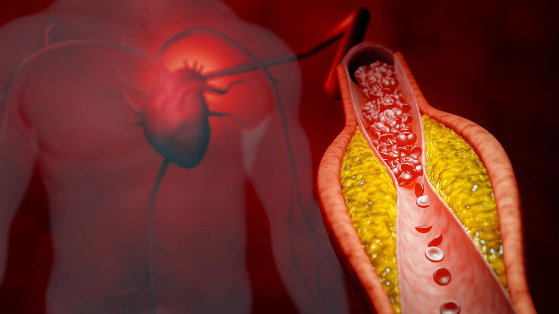
Platelets are vital in healing cuts, but in individuals with heart disease, they can form dangerous clots within arteries, leading to heart attacks and strokes.Currently, monitoring the effectiveness of antiplatelet drugs used to prevent thes clots is challenging. This new technology addresses that need.
The system utilizes a “frequency-division multiplexed (FDM) microscope” which captures thousands of images of blood cells flowing every second – akin to traffic cameras monitoring vehicles. AI then analyzes these images, identifying individual platelets, platelet clumps (indicating clotting risk), and other blood cells.
Testing on over 200 patients revealed a correlation between the number of platelet aggregates and the severity of acute coronary syndrome, demonstrating the technology’s ability to track clotting risk. Importantly, the researchers found that a standard blood draw from the arm provides comparable information to invasive procedures traditionally required to sample blood directly from the heart’s arteries.
“something as small as a blood cell can tell a big story about your health,” Zhou added.
This breakthrough offers the potential for personalized medicine, allowing doctors to tailor antiplatelet drug dosages based on an individual’s specific platelet behavior. As Dr. Hirose explains, patients respond differently to these medications, and this technology can help doctors “see how each individual’s platelets are behaving in real time” to optimize treatment and minimize risks like recurrent clots or bleeding.