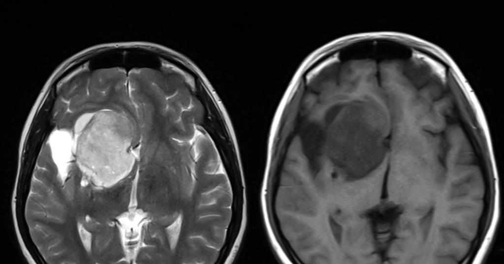
New Standard for Glioma Organoid Research Could Revolutionize Brain Cancer Treatment
A multi-institutional research team has established a standardized classification framework for glioma organoids, providing a rigorous methodology to improve the reproducibility and clinical relevance of brain cancer models. Published in the journal Nature Communications, this framework addresses long-standing inconsistencies in how laboratory-grown brain tumor models are characterized, potentially accelerating the path toward personalized precision oncology for glioblastoma patients.
Key Clinical Takeaways:
- The new framework standardizes the nomenclature and biological validation of glioma organoids, ensuring that lab models accurately reflect the genetic and phenotypic diversity of the original patient tumors.
- By improving model consistency, researchers can more reliably predict how specific tumor subtypes respond to experimental therapies, reducing the failure rate in translational drug development.
- This standardization facilitates a more uniform approach to high-throughput drug screening, moving the field away from non-standardized protocols that have historically hampered clinical translation.
Addressing the Reproducibility Crisis in Glioblastoma Research
Glioblastoma remains one of the most aggressive and treatment-resistant malignancies, with a median survival rate that has seen minimal improvement over the last two decades. The lack of reliable, standardized preclinical models is a primary bottleneck in developing effective pharmacotherapies. According to research led by teams at the University of Pennsylvania and published in Nature Communications, previous efforts to utilize glioma organoids—three-dimensional tissue cultures derived from patient samples—often suffered from “model drift,” where the organoid loses the molecular profile of the parent tumor over time.
The new framework, funded by the National Institutes of Health (NIH) and the National Cancer Institute (NCI), requires researchers to perform rigorous molecular characterization—specifically comparing the organoid’s genomic landscape to the original tumor biopsy. This ensures that the model maintains the pathogenesis and cellular heterogeneity essential for testing targeted inhibitors. Without such standards, researchers risk testing compounds against “off-target” cellular populations that no longer mirror the patient’s disease state, leading to high morbidity in early-phase clinical trial cohorts.
Biological Mechanism and Model Fidelity
The utility of this framework lies in its ability to bridge the gap between static 2D cell cultures and the complex, in vivo tumor microenvironment. As noted by Dr. H. Isaac Chen, a neurosurgeon and researcher at Penn Medicine, “These models allow us to see how tumors evolve in a controlled, three-dimensional space, but only if the model remains faithful to the patient’s biology.” The framework mandates clear documentation of the growth medium, extracellular matrix composition, and the duration of culture, all of which are critical variables that dictate the biological behavior of the model.
For clinicians managing high-grade glioma, the implication is a more refined pipeline for ex vivo drug sensitivity testing. By utilizing organoids that have been validated under these new parameters, oncology centers can theoretically perform “co-clinical trials,” testing various standard-of-care regimens against the patient’s own tumor organoids before initiating systemic treatment. Patients seeking advanced diagnostic or personalized treatment planning should consult with board-certified neuro-oncology specialists who are currently integrating these high-fidelity models into their research-backed clinical practice.
Clinical Triage and the Future of Translational Studies
The shift toward standardized organoid research necessitates a change in how clinical laboratories and pharmaceutical entities manage their research pipelines. Because these models now require standardized validation, institutions must invest in advanced genomic sequencing and high-resolution imaging to meet the new benchmark. For pharmaceutical firms and diagnostic developers, this represents a significant regulatory hurdle but ultimately offers a more stable environment for FDA/EMA drug approval filings.
Entities currently navigating the complexities of implementing these models within a clinical setting should seek guidance from specialized healthcare compliance and research consultants. Ensuring that your facility meets the latest standards for biological model validation is essential for maintaining integrity in translational studies. As the industry moves toward this standardized approach, the focus remains on reducing the gap between preclinical success and clinical efficacy.
The Evolving Landscape of Brain Cancer Therapeutics
The integration of these standardized models is expected to refine the selection process for Phase I and Phase II trials. By weeding out ineffective compounds using highly accurate 3D models before they reach human subjects, the medical community can better protect patients from the risks associated with experimental neuro-oncology treatments. The framework, as detailed in the recent Nature Communications study, provides the necessary criteria to ensure that every organoid used in research is a high-fidelity representation of the tumor it aims to treat.

Future research will likely focus on incorporating immune cells into these organoid models to better simulate the tumor-immune interface, a critical factor in the efficacy of modern checkpoint inhibitors and CAR-T cell therapies. As this methodology matures, it will remain a cornerstone for oncologists and researchers dedicated to improving the standard of care for patients with complex gliomas. For those currently managing a diagnosis, engaging with leading medical centers and oncology research hubs that utilize state-of-the-art diagnostic and experimental models is the most effective way to access emerging, evidence-based treatment strategies.
Disclaimer: The information provided in this article is for educational and scientific communication purposes only and does not constitute medical advice. Always consult with a qualified healthcare provider regarding any medical condition, diagnosis, or treatment plan.