New Electricity-Based Technique Could Replace LASIK for Vision Correction
Researchers are testing a non-invasive vision correction technique that reshapes the cornea using electricity instead of traditional lasers or surgical incisions, according to initial experimental results. Known as electromechanical reshaping, or EMR, the method applies a small electrical voltage to alter the chemical environment inside corneal tissue temporarily, increasing its flexibility for precise structural adjustments.
Key Findings on Electrical Corneal Reshaping
- Electromechanical reshaping uses platinum lenses as both electrical poles and physical molds to adjust corneal curvature without tissue ablation.
- Laboratory trials on 12 ex vivo rabbit eyes successfully altered the focusing power of 10 treated specimens simulating myopia.
- The entire reshaping process takes approximately one minute, functioning through a controlled pH shift that weakens tissue rigidity temporarily.
Mechanism of Action and Platinum Lens Application
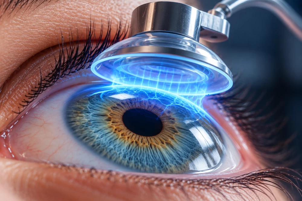
Unlike standard procedures such as LASIK, which permanently excise precise amounts of corneal tissue to alter its curvature, electromechanical reshaping modifies the existing architecture without removing any biological material. The procedure utilizes specialized platinum lenses placed directly over the cornea. These lenses serve a dual purpose, acting simultaneously as a physical mold for the targeted shape and as an active electrode.
When a low electrical voltage runs through the system, it induces a controlled, localized shift in pH within the cornea. This temporary chemical adjustment weakens specific molecular bonds responsible for the inherent rigidity of the tissue. Once softened, the cornea adapts to the contours of the platinum mold within roughly one minute. Removing the electrical stimulus restores normal physiological pH, allowing the tissue to lock into its newly formed geometry.
Experimental Validation in Preclinical Models
Initial testing of the electromechanical reshaping technique involved 12 rabbit eyes evaluated ex vivo. Investigators treated 10 of these eyes specifically to simulate myopia, or nearsightedness. Across all 10 treated specimens, the procedure successfully adjusted the corneal shape to match the required focusing power parameters.
Despite these promising laboratory metrics, the technique remains in an early preclinical phase. Researchers have not yet tested the procedure on human subjects. Current data is strictly limited to isolated animal tissue outside the body, leaving several questions regarding long-term biomechanical stability and cellular safety unanswered.
The development team plans to conduct further investigations to evaluate how well the cornea maintains its modified shape over extended periods and to confirm the safety of the surrounding cellular matrix. Because this approach avoids permanent tissue removal, investigators suggest it could eventually offer a simpler and less costly alternative to existing refractive surgeries, while bypassing certain risks associated with laser ablation.
Transitioning this experimental protocol toward human clinical trials will require extensive safety profiling. Patients considering vision correction options should consult with qualified ophthalmologists or optometrists to discuss established surgical and non-surgical treatments currently available under standard clinical care.
Disclaimer: The information provided in this article is for educational and scientific communication purposes only and does not constitute medical advice. Always consult with a qualified healthcare provider regarding any medical condition, diagnosis, or treatment plan.