Modern Radiotherapy Reduces Heart Disease Risk in Breast Cancer Patients
The clinical objective of breast cancer radiotherapy is clear: eliminate malignant cells while sparing healthy tissue. However, the heart’s proximity to the left breast creates a persistent risk of collateral radiation exposure, leading to a complex balance between oncological success and long-term cardiovascular morbidity.
Key Clinical Takeaways:
- Modern radiation techniques have significantly reduced the risk of major cardiac events compared to historical standards of care.
- The EARLY-HEART study reveals that even asymptomatic patients may develop subclinical changes in heart structure and function within two years of treatment.
- Advanced imaging, specifically cardiac MRI and LV GLS measurement, is essential for detecting early heart stress before clinical heart failure occurs.
For decades, the medical community has grappled with the pathogenesis of radiation-induced heart disease. Early landmark data, such as the study by Darby et al. Covering patients treated between 1973 and 2001, established a direct correlation between the indicate heart dose (MHD) and the probability of major cardiac events. This historical context created a climate of caution, as the incidence of ischemic heart disease remained a significant concern for breast cancer survivors. As radiotherapy evolved, the focus shifted toward minimizing the volumetric dose to the heart to prevent long-term cardiotoxicity.
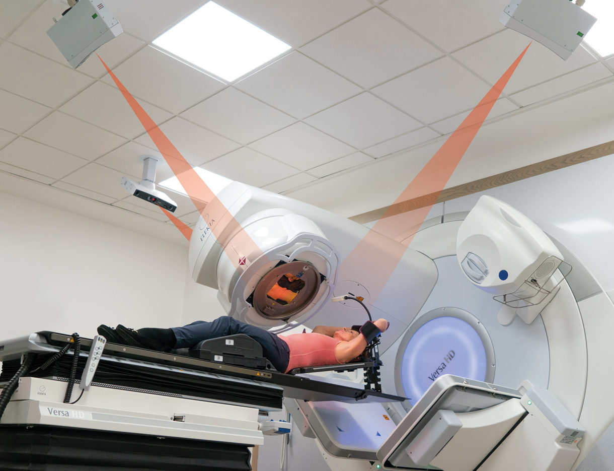
The evolution of external beam radiation therapy (EBRT) has largely succeeded in lowering the risk of overt heart failure. By employing deeper precision and lower doses, modern protocols aim to mitigate the risk of ischemic heart disease. Yet, the quest for “zero risk” is complicated by subclinical alterations that escape traditional diagnostic tools. This gap in detection is where current research, specifically the EARLY-HEART study, provides critical intelligence.
Decoding Subclinical Cardiac Stress: The EARLY-HEART Findings
The EARLY-HEART study, part of the European Union-funded Implications of Medical Low Dose Radiation Exposure (MEDIRAD) project, sought to identify the earliest markers of cardiac dysfunction. Conducted between December 2017 and September 2019, the research tracked 138 women across three European institutions. Unlike previous cohorts, these participants received lumpectomies and local radiation therapy without chemotherapy and were asymptomatic for cardiovascular disease at the start of the study.

Researchers utilized centralized cardiac MRI and automated strain measurement to monitor left ventricular global longitudinal strain (LV GLS). In the realm of cardiology, LV GLS is regarded as a highly sensitive early indicator of dysfunction, often predictive of future heart failure or heart attacks long before the ejection fraction drops. The results indicate that radiation exposure, even at modern levels, induces subtle but universal changes in heart architecture.
| Cardiac Metric | Baseline Status | Two-Year Follow-up Outcome |
|---|---|---|
| Heart Chamber Size | Normal | Slightly smaller left ventricular end-diastolic volume |
| Blood Output | Normal | Decreased stroke volume |
| Structural Integrity | Stable | Increase in cardiac remodeling |
| Clinical Symptoms | Asymptomatic | Remained asymptomatic |
The data suggests a paradoxical state: while the patients remain clinically healthy and asymptomatic, their hearts are undergoing structural remodeling. This subclinical decline—characterized by smaller chambers and reduced blood output—highlights a critical window for intervention. For patients navigating this recovery phase, the transition from oncological care to cardiovascular surveillance is paramount. It is highly recommended to coordinate with board-certified cardiologists specializing in cardio-oncology to monitor these structural shifts.
From Mean Heart Dose to Personalized Surveillance
The shift from the broad dose-response models seen in The New England Journal of Medicine to the precise imaging seen in RSNA reports marks a new era of personalized care. We are moving away from a “one size fits all” radiation approach toward a model where the specific cardiac dose received by a patient informs their lifelong screening schedule.
The morbidity associated with radiotherapy is not uniform. Those who receive higher cardiac doses are more susceptible to the remodeling observed in the MEDIRAD project. This necessitates a multidisciplinary approach to treatment planning. To minimize these risks at the outset, patients should ensure their treatment is mapped by expert radiation oncologists using the latest dose-reduction technologies.
The detection of these changes relies heavily on the availability of high-resolution imaging. Standard echocardiograms may miss the subtle decreases in stroke volume or the nuanced changes in LV GLS. There is a growing clinical demand for advanced diagnostic imaging centers capable of performing validated automated strain measurements and centralized cardiac MRI.
The Future of Cardio-Oncology Integration
The trajectory of breast cancer treatment is moving toward a symbiotic relationship between oncology and cardiology. The fact that all participants in the EARLY-HEART study remained asymptomatic despite structural changes suggests that early detection allows for proactive management. By identifying “heart stress” in its subclinical phase, clinicians can potentially implement lifestyle interventions or pharmacological protections to prevent the progression toward ischemic heart disease.
The evidence from the MEDIRAD project underscores that while modern radiotherapy has drastically cut the risk of catastrophic heart failure, it has not entirely eliminated the biological impact on the myocardium. The focus must now shift to long-term, data-driven surveillance. The goal is no longer just survival, but the preservation of cardiovascular longevity.
As we refine the standard of care, the integration of cardiac monitoring into the breast cancer survivorship plan will become mandatory. Patients and providers should prioritize a network of vetted specialists to ensure that the victory over cancer is not offset by a late-stage cardiovascular event. Finding a coordinated care team through our directory ensures that these sophisticated monitoring protocols are implemented with precision.
Disclaimer: The information provided in this article is for educational and scientific communication purposes only and does not constitute medical advice. Always consult with a qualified healthcare provider regarding any medical condition, diagnosis, or treatment plan.