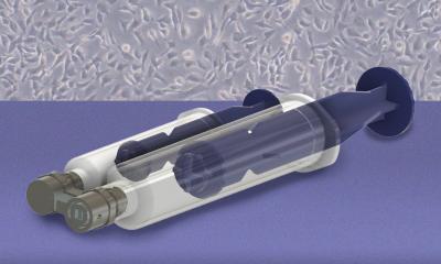
MIT Researchers Develop New Device for Early Ovarian Cancer Detection
Researchers at the Massachusetts Institute of Technology and Johns Hopkins University developed a handheld microfluidic device to gently collect living cells from specific regions of surgically removed fallopian tubes, aiming to improve the early detection of high-grade serous ovarian cancer.
Engineering a Novel Microfluidic Tool
The technology addresses a critical clinical bottleneck by allowing pathologists to study living cellular behavior rather than relying solely on preserved, sectioned tissue. The handheld microfluidic instrument uses a vacuum seal to isolate tiny tissue areas and applies controlled fluid shear stress to detach viable cells without destroying surrounding structures.
Recovered cells remain viable and successfully grow in culture, enabling downstream applications like organoid creation and personalized drug response testing. Funding for the interdisciplinary effort comes from Break Through Cancer, connecting engineering expertise from MIT with gynecologic oncology insights from Johns Hopkins.

Overcoming Pathological Workflows
The standard pathological workflow for examining fallopian tubes involves placing freshly excised tissue into a chemical preservative before slicing it into microscopic sections. While this preserves physical architecture, it kills the cells, rendering them incapable of growth or functional analysis.
Seeking an alternative, MIT mechanical engineering professor Kripa Varanasi collaborated with Johns Hopkins gynecologic oncologist Rebecca Stone after an operating room visit inspired the team. The researchers realized that fluid flow could generate precise shear stress capable of lifting loose cells off tissue surfaces.
Precision Harvesting in the Laboratory
The resulting 3D-printed device uses a dual-syringe mechanism. The first syringe establishes a vacuum seal against the target tissue to prevent leakage, while the second pushes liquid through a localized microfluidic channel.
Laboratory testing demonstrated that adjusting the fluid shear stress successfully detached cells while maintaining high cellular viability compared to conventional approaches. The team tested the system on fresh human fallopian tube samples shipped directly from Johns Hopkins, recovering cells capable of thriving in culture.
Advancing Personalized Cancer Care
Discoveries over the past two decades established that many high-grade serous ovarian cancers originate within the fallopian tubes. Catching these precursor lesions remains exceptionally difficult because they are often microscopic.
While preventive removal of fallopian tubes after childbearing years helps patients at increased risk, diagnosing the earliest stages of disease in retained tissue requires new diagnostic modalities. By capturing living cells from specific anatomical sites, researchers can construct organoids and disease models. These living models pave the way for testing how an individual patient’s cells respond to different therapeutic agents, ultimately supporting the development of personalized cancer care strategies.
Disclaimer: The information provided in this article is for educational and scientific communication purposes only and does not constitute medical advice. Always consult with a qualified healthcare provider regarding any medical condition, diagnosis, or treatment plan.