Immediate Neoumbilicoplasty After Symptomatic Umbilical Lipoma Excision: A Case Report
Surgical management of symptomatic umbilical lipomas frequently presents a cosmetic challenge, as traditional excision often results in a flattened or distorted navel. A recent case report published in Cureus demonstrates that immediate neoumbilicoplasty—utilizing local tissue rearrangement and fascial fixation—can effectively excise the mass while restoring the natural aesthetic contour of the umbilicus.
Key Clinical Takeaways:
- Immediate neoumbilicoplasty allows for the simultaneous removal of umbilical lipomas and the preservation or reconstruction of the umbilical shape.
- Fascial fixation is a critical step in preventing umbilical flattening, providing a stable anchor to recreate the central depression.
- This technique offers a reproducible, low-morbidity alternative to simple excision, which often leaves the patient with a suboptimal umbilical appearance.
Clinical Context and Pathogenesis of Umbilical Lipomas
Umbilical lipomas are benign adipose tumors that, while relatively uncommon, can cause significant patient discomfort, local tenderness, and aesthetic concern. The umbilicus is a complex anatomical structure, and its disruption during the excision of a lipoma often leads to a “flat” or “obliterated” navel. According to the report in Cureus, standard surgical approaches have historically prioritized the removal of the pathology over the structural integrity of the umbilical depression. This creates a clinical gap where patients may be cured of their physical symptoms but dissatisfied with the resulting abdominal contour.
The pathogenesis of these lesions involves the proliferation of mature adipocytes within the umbilical ring or the surrounding subcutaneous tissue. When a lipoma exceeds a certain volume, it exerts pressure on the overlying skin and the underlying fascia, leading to a visible protrusion. Surgical intervention requires a delicate balance: the surgeon must achieve complete resection of the encapsulated fatty tissue while ensuring that the deep umbilical attachment is maintained or recreated to preserve the natural “in-y” morphology.
Surgical Technique: Local Tissue Rearrangement and Fascial Fixation
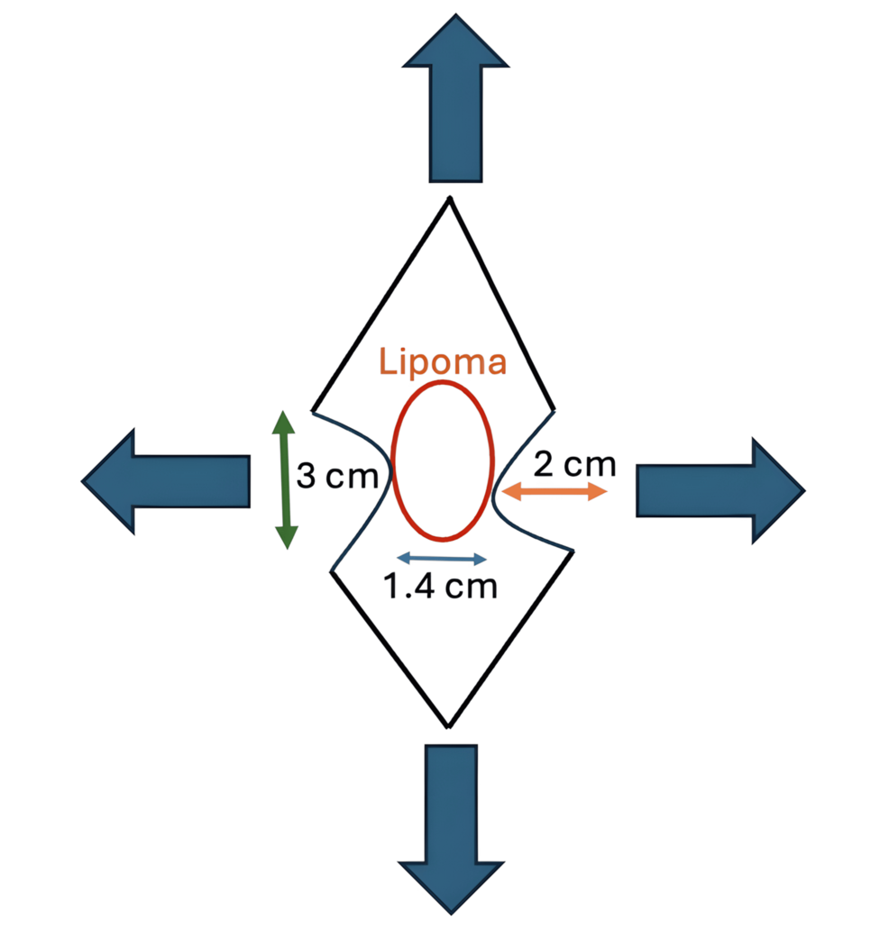
The procedure detailed in the Cureus case report centers on a refined surgical approach that minimizes tissue trauma. After the excision of the lipoma, the surgeon performs local tissue rearrangement—utilizing the redundant skin left behind by the mass—to recreate the umbilical hood and depth. A pivotal component of this technique is fascial fixation, where the deep dermis of the new umbilicus is sutured directly to the rectus abdominis fascia.
This fixation mimics the natural anatomical tethering of the umbilicus. By securing the base of the umbilicus to the fascia, the surgeon prevents the postoperative flattening that typically follows the excision of significant umbilical masses. This approach is consistent with broader plastic surgery principles where the restoration of anatomical “anchors” is essential for functional and aesthetic outcomes. Patients considering this procedure should consult with board-certified plastic surgeons who specialize in umbilical reconstruction to ensure the highest standard of care.
Evaluating Clinical Outcomes and Morbidity
The patient in the Cureus report experienced an uncomplicated recovery, with the neoumbilicoplasty yielding a natural-appearing umbilicus. While this is an N-of-1 case study, it contributes to the growing body of literature suggesting that immediate reconstruction is superior to delayed or secondary revision. The morbidity associated with this technique is minimal, primarily involving standard risks associated with minor soft tissue surgery, such as minor hematoma or seroma formation, which are mitigated through careful surgical technique and proper postoperative compression.
For individuals experiencing persistent pain or discomfort in the umbilical region, a differential diagnosis is essential. These symptoms can sometimes mimic umbilical hernias, urachal remnants, or endometriomas. It is highly recommended to seek an evaluation from a specialized diagnostic center or a general surgeon to accurately characterize the mass via imaging, such as an ultrasound or MRI, before proceeding with elective excision. Accurate preoperative assessment ensures that the surgical plan addresses both the lesion and the potential for umbilical wall weakness.
Future Directions in Umbilical Reconstruction
As surgical techniques continue to evolve, the focus is shifting toward “scarless” or “minimally invasive” aesthetic outcomes. The integration of neoumbilicoplasty during primary excision represents a move toward comprehensive care that values patient quality of life alongside clinical resolution. Future research may involve larger cohorts to standardize these rearrangement techniques across different patient demographics and body mass indices.
Healthcare providers, including surgical clinics and outpatient centers, should note the importance of incorporating aesthetic considerations into routine minor surgeries. Patients seeking to address umbilical abnormalities should engage with vetted healthcare providers who prioritize both the functional resolution of the lipoma and the cosmetic restoration of the navel. By aligning clinical practice with these refined reconstructive techniques, providers can improve patient satisfaction and reduce the demand for secondary revisional procedures.
Disclaimer: The information provided in this article is for educational and scientific communication purposes only and does not constitute medical advice. Always consult with a qualified healthcare provider regarding any medical condition, diagnosis, or treatment plan.