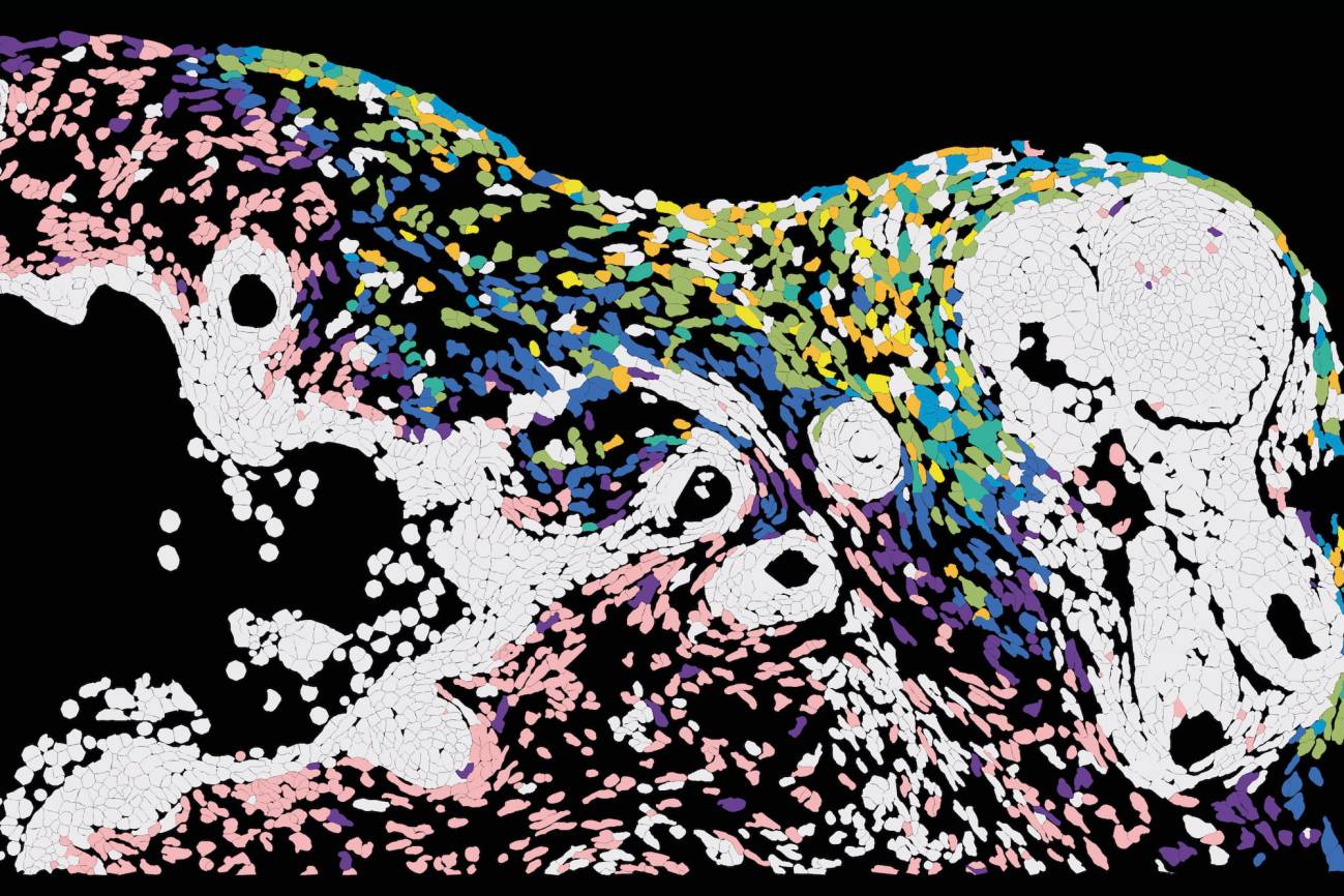
How Herpes Simplex Virus 1 Spreads Through Human Skin Organoids
Researchers in Germany have mapped how the herpes simplex virus type 1 moves through human skin layers using three-dimensional tissue models, agenciasinc.es reported on October 30, 2026.
There is a possible relationship between the persistent presence of HSV-1 and processes linked with Alzheimer’s disease, though that link continues under investigation.
- Dermal fibroblasts accumulated high viral loads early in the infection cycle, whereas basal keratinocytes and hair follicle cells showed resistance and delayed viral entry.
- Combining tissue imaging with spatial transcriptomics revealed coordinated activation of TNF signaling molecules.
Skin Organoids Reveal Unequal Cellular Susceptibility
Human skin features a multi-layered structure consisting of an outer epidermis packed with keratinocytes, a supportive dermis containing fibroblasts, and hair follicles interlaced with sensory nerve fibers. To capture this complexity, researchers built hair-bearing skin organoids. As Emanuel Wyler of the Max Delbrück Center explained to agenciasinc.es, these three-dimensional cultures reconstruct native tissue architecture with various cell types, though they lack immune cells and blood vessels.
When tracking the pathogen across these models, the team observed that the virus did not infect every tissue component equally. According to findings highlighted by Infobae, dermal fibroblasts stored large amounts of viral material during early infection phases. Conversely, basal keratinocytes at the lower edge of the epidermis and multiple cell types within the hair follicles contained significantly lower viral loads, even when adjacent cells were heavily infected. This disparity indicates that distinct cell types provide varying conditions for viral entry and replication.

Spatial Transcriptomics Map Cellular Defense Responses
To examine the active infection front without disrupting tissue architecture, researchers paired high-resolution imaging with spatial transcriptomics. This method measures gene activity within individual cells while preserving their exact anatomical location.
Members of the tumor necrosis factor signaling family activated in a spatially coordinated manner. Emanuel Wyler noted that these TNF molecules stimulate the immune system and likely dictate how effectively the body controls an infection.

Future Models Await Immune Cells and Blood Vessels
The current organoid models demonstrate how HSV-1 moves through structured layers, but the absence of a vascular network and immune cells leaves open questions regarding how the human body halts the pathogen before it reaches sensory nerves. Future research initiatives aim to integrate immune cells and blood vessels into advanced organoid designs to clarify the precise mechanisms that allow certain cells to suppress viral replication while others sustain it.
Disclaimer: The information provided in this article is for educational and scientific communication purposes only and does not constitute medical advice. Always consult with a qualified healthcare provider regarding any medical condition, diagnosis, or treatment plan.