Extracardiac Mediastinal Mass Presenting With Cardiac Arrhythmias: A Case Report
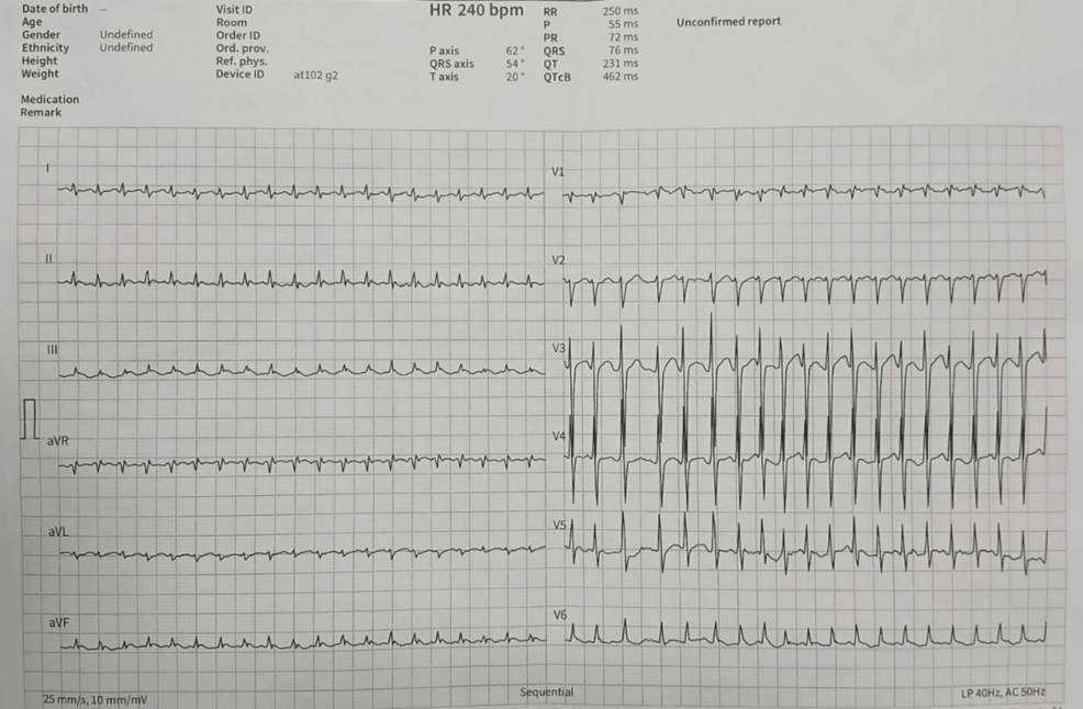
An extracardiac mediastinal mass can directly precipitate acute cardiac arrhythmias by exerting mechanical pressure on underlying myocardial structures, according to a recent clinical case report published in Cureus. Physicians diagnosing unexplained rhythm disturbances must consider thoracic space-occupying lesions in differential workups, as physical compression of cardiac chambers disrupts normal electrophysiological pathways.
Key Clinical Takeaways:
- Mediastinal masses located outside the heart can mechanically irritate myocardial tissue and trigger symptomatic cardiac arrhythmias.
- Standard diagnostic evaluations for refractory rhythm disorders should include comprehensive thoracic imaging to rule out extracardiac structural compression.
- Multidisciplinary surgical and electrophysiological management remains necessary when space-occupying chest lesions compromise hemodynamic stability.
Pathogenesis and Electrophysiological Disruptions
The biological mechanism linking a mediastinal mass to a cardiac arrhythmia involves direct mechanical stretch and irritation of the atrial or ventricular myocardium. When a tumor or cyst enlarges within the confined mediastinal space, it compromises cardiac compliance. This physical constraint alters local action potential durations and refractory periods across the affected tissue. According to findings detailed in the medical literature via PubMed Central, localized pressure gradients can provoke ectopic pacemaker activity, leading to premature beats or sustained tachyarrhythmias. Clinicians evaluating patients with new-onset palpitations or syncope must look beyond primary cardiac etiologies and assess the mediastinum.
Diagnostic Triage and Imaging Protocols
Isolating the root cause of compression-induced arrhythmias requires advanced diagnostic imaging modalities, including contrast-enhanced computed tomography and magnetic resonance imaging of the chest. These scans map the precise anatomical relationship between the extracardiac mass and adjacent cardiac chambers. For patients exhibiting persistent rhythm instability unresponsive to standard antiarrhythmic pharmacotherapy, timely surgical evaluation is critical. It is highly recommended to consult with vetted board-certified cardiologists and cardiac electrophysiologists to map aberrant conduction pathways. Simultaneously, coordinating with specialized thoracic surgery centers ensures that structural decompression can be executed safely before irreversible myocardial damage occurs.
Future Trajectory in Managing Complex Thoracic Pathology
As diagnostic imaging resolution improves, identifying small mediastinal masses before they induce severe hemodynamic compromise will become more feasible. Integrating advanced electrophysiological mapping with real-time structural imaging allows surgical teams to target interventions precisely. Mitigating long-term morbidity in these complex presentations ultimately depends on early cross-specialty collaboration between oncologists, surgeons, and rhythm specialists.
Disclaimer: The information provided in this article is for educational and scientific communication purposes only and does not constitute medical advice. Always consult with a qualified healthcare provider regarding any medical condition, diagnosis, or treatment plan.