
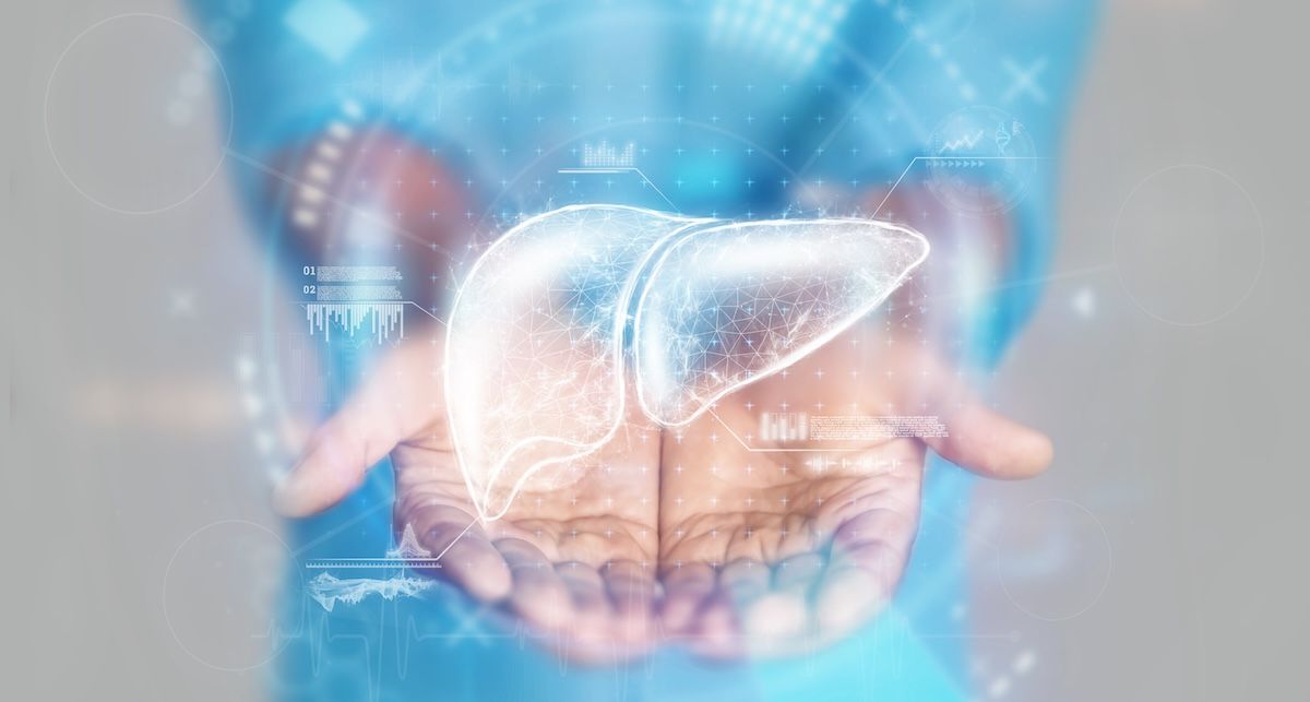
Unraveling the Spatial Landscape of Metabolic Dysfunction-Associated Steatohepatitis (MASH)
Metabolic dysfunction-associated steatohepatitis (MASH), formerly known as nonalcoholic steatohepatitis (NASH), is a growing global health concern. This serious liver condition, characterized by inflammation and fibrosis, can progress to cirrhosis and liver cancer. Recent research published in Nature Genetics1 has unveiled a groundbreaking spatially resolved, multi-omics atlas of human MASH, offering unprecedented insights into the cellular interactions and molecular mechanisms driving disease progression. This detailed mapping promises to revolutionize our understanding of MASH and pave the way for more targeted and effective therapies.
The Evolution of Understanding Liver Disease: From NASH to MASH
For years, nonalcoholic fatty liver disease (NAFLD) was an umbrella term encompassing a range of liver conditions linked to metabolic dysfunction. However, it became increasingly clear that not all cases of fatty liver were created equal. The term MASH was adopted to specifically denote the more aggressive form of the disease characterized by inflammation and cellular damage. This shift in terminology reflects a growing understanding of the complex interplay between metabolic factors, immune responses, and genetic predisposition in the advancement of liver disease.
A New Dimension: The Power of Spatial Multi-Omics
Traditionally,studies of MASH have relied on analyzing bulk tissue samples,providing an average picture of cellular activity.However, this approach obscures the critical spatial institution of cells and molecules within the liver. the liver isn’t a homogenous organ; its function is highly organized,with metabolic and immune processes varying across different zones.The recent study overcomes this limitation by employing a suite of cutting-edge technologies – single-cell RNA sequencing, spatial transcriptomics, and spatial metabolomics – to create a detailed map of the liver’s microenvironment in health and disease.
- Single-cell RNA sequencing: Identifies the gene expression profiles of individual cells, revealing the diversity of cell types and their functional states.
- Spatial transcriptomics: Maps gene expression patterns within the tissue, preserving the spatial context of cells.
- Spatial metabolomics: Identifies the distribution of metabolites (small molecules involved in metabolism) within the tissue, providing insights into metabolic activity.
By integrating these datasets, researchers were able to create a comprehensive “atlas” of the MASH liver, revealing how cells interact and how metabolic pathways are altered in different regions of the organ.
Key Findings: Lipid-Associated Macrophages and the role of MITF
One of the most significant findings of the study was the identification of lipid-associated macrophages (LAMs) as key players in MASH progression. LAMs are a type of immune cell that accumulates in the fatty liver and plays a role in inflammation and fibrosis. The researchers found that LAMs were more abundant in individuals with MASH compared to those with simpler steatosis (fat accumulation without inflammation). Importantly, spatial analysis revealed that LAMs were concentrated in the pericentral regions of the liver, areas particularly vulnerable to metabolic stress and hypoxia (low oxygen levels).
Further examination pinpointed microphthalmia-associated transcription factor (MITF) as a crucial regulator of LAM function. MITF is a protein that controls gene expression, and the study showed that its activity was substantially elevated in LAMs from MASH patients. Increasing MITF levels in laboratory experiments enhanced the ability of macrophages to process lipids, suggesting that this transcription factor plays a key role in shaping the lipid-handling capabilities of these immune cells.
Unveiling the Fibrotic Niche
Fibrosis, the formation of scar tissue, is a hallmark of advanced MASH and a major driver of disease progression. The study’s spatial analysis revealed that fibrosis doesn’t occur uniformly throughout the liver but rather develops in localized “niches.” These niches are characterized by coordinated signaling between central vein endothelial cells (cells lining the central veins) and hepatic stellate cells (cells responsible for producing collagen, a key component of scar tissue). This finding suggests that targeting these localized fibrotic signals could be a promising therapeutic strategy.
Metabolomic Insights: Phospholipid Accumulation
The researchers also used spatial metabolomics to identify specific metabolic changes associated with MASH.they found an accumulation of phospholipids, a type of fat molecule, in the livers of MASH patients. This accumulation was linked to altered phospholipid metabolism mediated by LAMs, specifically involving pathways related to lipoprotein-associated phospholipase A2.These findings suggest that modulating phospholipid metabolism could be a potential therapeutic target.
Implications for Future Therapies
This spatially resolved multi-omics atlas represents a significant advance in our understanding of MASH. By providing a detailed map of the disease’s cellular and molecular landscape,it opens up new avenues for therapeutic intervention. Specifically,the findings suggest that targeting LAMs,MITF,or the localized fibrotic niches could be effective strategies for slowing or reversing disease progression.
While the study has limitations – including a relatively modest sample size and a cross-sectional design that doesn’t capture the dynamic changes of disease progression – it provides a valuable resource for researchers developing new therapies. Future studies will need to validate these findings in larger cohorts and explore the potential of targeting these pathways in clinical trials.
Looking Ahead: Personalized Medicine for MASH
The era of “one-size-fits-all” medicine is waning. Increasingly, treatment strategies are being tailored to the individual characteristics of each patient. The spatially resolved multi-omics data generated by this study could be instrumental in developing personalized approaches to MASH management. By analyzing a patient’s liver tissue at the single-cell and spatial level,clinicians could identify the specific pathways that are driving disease progression and select the most appropriate therapy.
Frequently Asked Questions (FAQ)
- What is MASH? MASH, or metabolic dysfunction-associated steatohepatitis, is a severe form of nonalcoholic fatty liver disease characterized by inflammation and fibrosis.
- Why is spatial resolution vital in studying MASH? The liver is a spatially organized organ, and understanding how cells interact within specific regions is crucial for understanding disease mechanisms.
- What are lipid-associated macrophages (LAMs)? LAMs are a type of immune cell that accumulates in the fatty liver and contributes to inflammation and fibrosis.
- What is MITF and how is it involved in MASH? MITF is a transcription factor that regulates lipid metabolism in LAMs and is elevated in MASH patients.
- What are the potential therapeutic implications of this research? Targeting LAMs, MITF, or localized fibrotic niches could be promising strategies for treating MASH.
References
1. Li Z, Luo G, Gan C, et al. Spatially resolved multi-omics of human metabolic dysfunction-associated steatotic liver disease. Nat Genet. 2025;57(12):3112-3125. doi:10.1038/s41588-025-02407-8
2. Metabolic dysfunction-associated steatotic liver disease (MASLD). Children’s Hospital of Philadelphia. Accessed January 16, 2026. https://www.chop.edu/conditions-diseases/metabolic-dysfunction-associated-steatotic-liver-disease-masld