Depression Linked to Shrinkage in Key Brain Memory Center
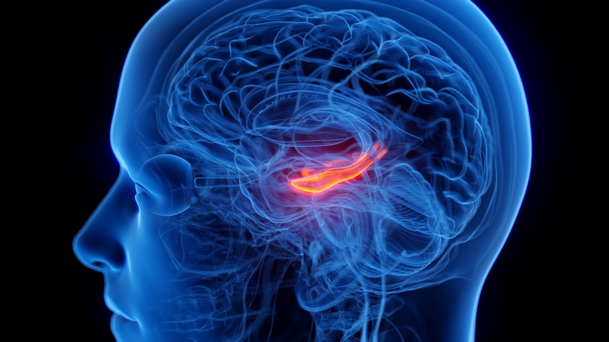
Researchers at the University of Southern California (USC) have identified a localized reduction in the volume of the CA23DG subregion of the hippocampus in individuals suffering from depression. Published in the journal Translational Psychiatry in 2026, the study suggests that this specific structural change—distinct from broader hippocampal atrophy—may represent a biological marker for depression that operates independently of known Alzheimer’s disease pathology.
Key Clinical Takeaways:
- A specific subregion of the hippocampus, known as CA23DG, appears smaller in patients with depression compared to those without the condition.
- The structural difference remains significant even after accounting for established Alzheimer’s risk factors, including amyloid and tau protein levels and the APOE ε4 genetic variant.
- The study suggests that the duration of depression, rather than just symptom severity, may be a primary driver of these hippocampal structural changes.
Defining the Hippocampal Pathogenesis
The hippocampus serves as the brain’s primary hub for memory consolidation, spatial navigation, and learning. While previous clinical literature has established a correlation between clinical depression and an elevated risk of cognitive decline, the precise neurological mechanisms have remained elusive. The USC team, utilizing MRI and PET imaging data from 2,009 cognitively unimpaired adults aged 50 to 90, focused on sub-segmenting the hippocampus to move beyond gross volumetric measurements.
The researchers specifically analyzed three distinct areas: CA1, the subiculum, and the composite region CA23DG, which includes the CA2 and CA3 fields along with the dentate gyrus. According to the findings, the volume reduction was isolated to the CA23DG region. This specificity is clinically relevant, as CA23DG is responsible for pattern separation—the process by which the brain distinguishes between similar memories—and the reconstruction of memories from partial cues.
“By looking closely at the individual parts of the hippocampus, we identified a specific area that may be particularly sensitive to depression in older adults,” stated lead author Danielle Luu, a doctoral student at the USC Mark and Mary Stevens Neuroimaging and Informatics Institute. The study, which was supported by institutional research funding, suggests that failing to differentiate between these subregions in prior research may have obscured the localized nature of these neuroanatomical changes.
Differential Diagnosis and Comorbidity
A critical component of this investigation involved determining whether the observed shrinkage was an early indicator of neurodegenerative disease. By adjusting for amyloid-beta and tau protein loads—the biological hallmarks of Alzheimer’s disease—the researchers found the association between depression and CA23DG volume persisted. This indicates that the structural alterations linked to depression are likely a distinct phenomenon from the amyloid-driven pathology seen in Alzheimer’s.
For patients and clinicians, these findings emphasize the importance of early intervention in mood disorders to mitigate long-term structural brain changes. Individuals experiencing persistent cognitive fog or memory concerns alongside mood symptoms should seek evaluation from a qualified healthcare provider to differentiate between functional depression-related changes and early-stage neurodegeneration.
The Role of Pharmacological Intervention
The study introduced a complex variable regarding antidepressant usage. Researchers observed that participants taking antidepressants demonstrated reduced volume in both the CA23DG and CA1 regions. The authors noted that this finding is counterintuitive, as pharmacological treatment is intended to stabilize mood and potentially shield the brain from the chronic stress-related damage associated with untreated depression.

The research team posited a “severity bias” hypothesis: patients with more severe or treatment-resistant depression are more likely to be prescribed medication, meaning the medication use may be a proxy for the intensity of the underlying depressive state rather than the cause of the atrophy. Furthermore, data indicated that the duration of the depressive state correlated more strongly with volume loss than recent symptom scores, suggesting that chronic, low-grade depression may exert a cumulative, deleterious effect on hippocampal architecture.
Clinical management of such cases requires a nuanced approach. Patients may benefit from a comprehensive review of their neuro-psychiatric care plan with a qualified healthcare provider to ensure that both symptom management and long-term neuro-protection are being addressed. As clinical understanding of these biomarkers evolves, the ability to monitor hippocampal sub-volumes may eventually inform more personalized treatment protocols for aging populations.
Future Research Trajectories
While the study provides a significant advancement in mapping the biological footprint of depression, it remains an observational analysis. Future longitudinal trials are required to establish whether these structural changes are reversible or if they represent a permanent shift in neural circuitry. Continued research into the interactions between neuro-inflammation, chronic stress, and hippocampal subfield integrity will be essential for developing targeted therapies that move beyond general symptom relief.

Disclaimer: The information provided in this article is for educational and scientific communication purposes only and does not constitute medical advice. Always consult with a qualified healthcare provider regarding any medical condition, diagnosis, or treatment plan.