
“`html
Hypertrophic Cardiomyopathy: Understanding the Genetic Heart Disease
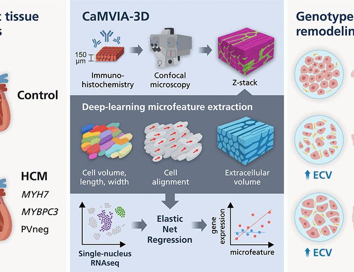
Hypertrophic cardiomyopathy (HCM) is a genetic heart condition characterized by the thickening of the heart muscle, particularly the left ventricle. This thickening can obstruct blood flow and lead to a range of complications, including sudden cardiac death. While HCM affects individuals of all ages, it’s a leading cause of unexpected death in young athletes and remains a significant concern globally. recent research is focusing on the complex three-dimensional organization of cardiac tissue to better understand and treat this disease.
What is Hypertrophic Cardiomyopathy?
At its core,HCM isn’t about the heart getting bigger in the way it does with high blood pressure. instead,the heart muscle,specifically the left ventricle (the main pumping chamber),thickens abnormally. This thickening can occur in various patterns and locations within the heart. The thickening often reduces the space available for blood, making it harder for the heart to pump efficiently. In certain specific cases, the thickened muscle obstructs the outflow of blood, a condition known as obstructive HCM.
causes and genetics
HCM is primarily caused by mutations in genes that control the production of heart muscle proteins. These mutations are often inherited, meaning they are passed down from parents to children. However, not everyone who inherits a gene mutation will develop HCM; the condition exhibits variable expressivity and incomplete penetrance. This means the severity of the disease can differ greatly among family members, and some individuals with the gene mutation may never show symptoms.
Over a dozen different genes have been linked to HCM, with mutations in the MYH7 gene (encoding beta-myosin heavy chain) being the most common. The National Library of Medicine provides detailed information on the genetic basis of HCM.
Symptoms of HCM
Many people with HCM have no symptoms for years, or even a lifetime. When symptoms do occur, they can vary widely in severity and may include:
- Shortness of breath, especially during exercise
- Chest pain, often mistaken for angina
- Dizziness or lightheadedness
- Fainting (syncope), particularly during or after exercise
- Palpitations (feeling of rapid, fluttering, or pounding heartbeat)
- Fatigue
- Sudden cardiac arrest
symptoms often worsen with physical activity. It’s crucial to note that symptoms can mimic other, less serious conditions, making diagnosis challenging.
Diagnosis
diagnosing HCM typically involves a combination of tests:
- Echocardiogram: This is the primary diagnostic tool, using sound waves to create images of the heart, revealing the thickness of the heart muscle and any obstructions.
- Electrocardiogram (ECG): This test measures the electrical activity of the heart and can detect abnormal rhythms.
- Cardiac MRI: Provides detailed images of the heart muscle and can definitely help differentiate HCM from other conditions.
- Genetic Testing: Can identify specific gene mutations associated with HCM, aiding in diagnosis and family screening.
- Cardiac Catheterization: In some cases, this invasive procedure may be used to measure pressures within the heart chambers.
Treatment Options
Treatment for HCM aims to relieve symptoms, prevent complications, and reduce the risk of sudden cardiac death. Treatment options include:
- Medications: Beta-blockers and calcium channel blockers can help slow the heart rate and reduce the force of heart contractions, improving blood flow and relieving symptoms. Disopyramide is sometimes used to reduce obstruction.
- Implantable Cardioverter-Defibrillator (ICD): Recommended for individuals at high risk of sudden cardiac death,an ICD monitors heart rhythm and delivers an electrical shock to restore a normal rhythm if a dangerous arrhythmia occurs. The American Heart Association provides guidelines on ICD implantation.
- Septal Myectomy: A surgical procedure to remove a portion of the thickened septum (the wall between the ventricles) to relieve obstruction.
- alcohol Septal Ablation: A less invasive procedure where alcohol is injected into a small artery supplying the thickened septum, causing it to shrink.
Recent Advances and Research
Current research is delving into the