The study found that the reduction of epithelial pigment-derived protein aspect is a determinant of age-related alterations in the retina.
–
Mice with no the protective protein in their eyes demonstrate indicators identical to age-related macular degeneration.
Loss of the protein pigment-derived issue (PEDF), which guards the supportive cells of the retina, may well endorse age-related variations in the retina, in accordance to a current National Eye Institute (NEI) examine in mice.
Age-linked health conditions of the retina, these types of as age-connected macular degeneration (AMD), can lead to blindness because the retina is the mild-sensitive tissue at the back again of the eye. The new facts could aid the growth of medication to quit AMD and other retinal getting old problems. The study was revealed in Intercontinental Journal of Molecular Sciences. NEI is component of the Countrywide Institutes of Wellness.
“Folks have termed PEDF the ‘youth’ protein due to the fact it is plentiful in the young retina, but decreases with age,” mentioned Patricia Becerra, PhD, president of the protein construction and functionality division at the Countrywide Institute. investigate scientist and direct creator of the analyze. “This study exhibits for the initially time that a very simple elimination of PEDF results in a sequence of genetic alterations that mimic retinal growing older.”
The retina is created up of levels of cells that operate alongside one another to recognize and interpret light alerts, which the mind takes advantage of to create sight. The photosensitive photoreceptors in the retina are found at the major of a supporting cell layer referred to as the retinal pigment epithelium (RPE). When photoreceptors detect light, the RPE feeds them and recycles their “outer parts,” which burn up and cut off the strategies anytime the receptors detect mild.
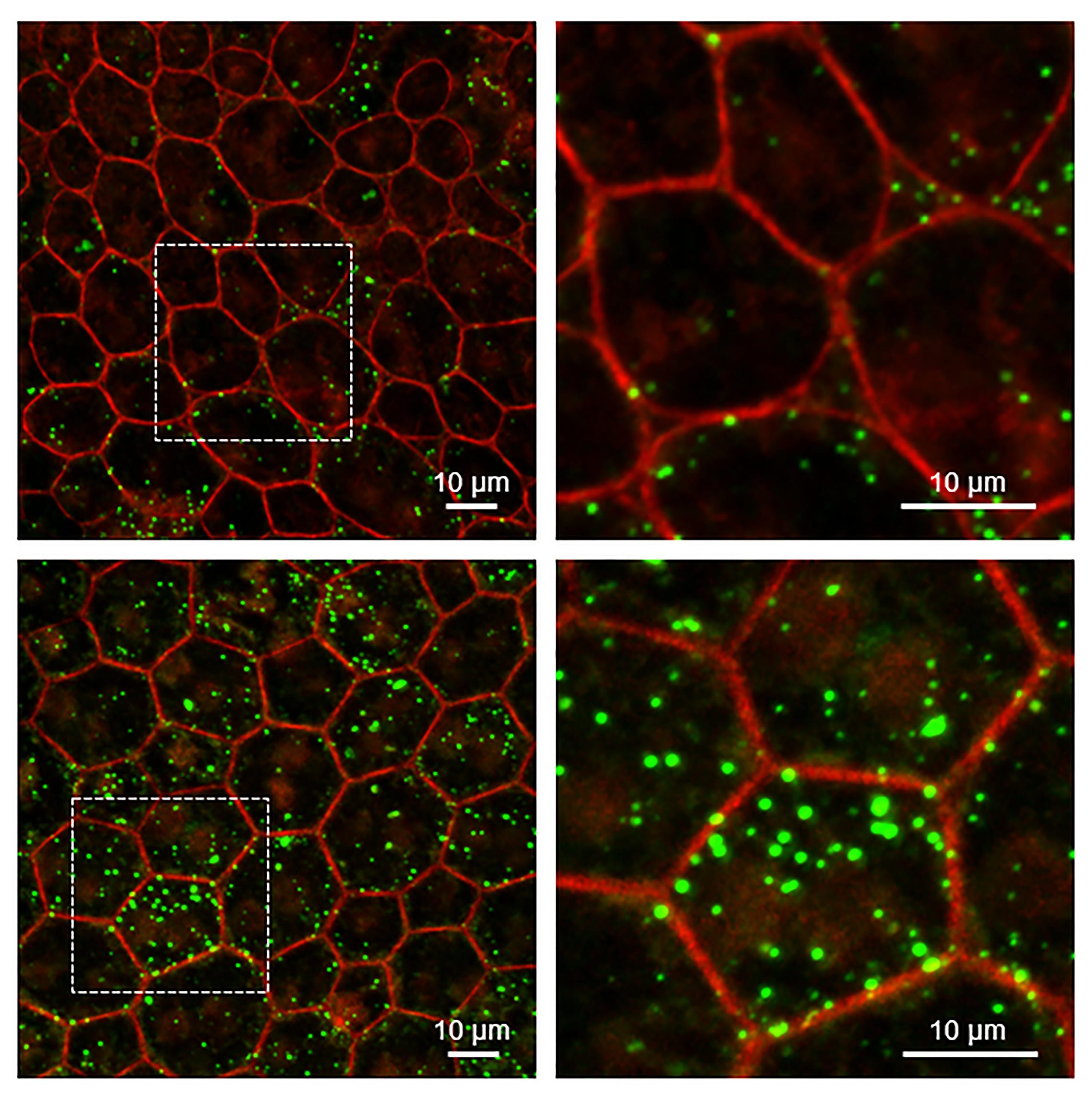
RPEs from mice without the need of Serpin1 accumulate a lot more lipids than wild-kind mice. Really large resolution microscopy of tissues from wild-type (upper) and Serpin1-null (reduce) RPE mice. The comprehensive pictures on the suitable are enlarged locations of the RPE cloth depicted on the remaining (dashed sq. spot). The edges of the RPE cells are coloured crimson and the amassed lipids are coloured environmentally friendly. Credit history: Ivan Ripostini, NEI
–
Photoreceptor cells reduce the capability to produce new segments and so lose the skill to detect light if the RPE is unable to provide them with recycled components from the ends of the previous outer section. With out the vitamins furnished by the RPE, the photoreceptors would die. Getting older (senescence) or the death of RPE cells in the retina prospects to eyesight reduction in folks with AMD or sure types of retinal atrophy.
Former exploration by Becerra and other groups has proven that PEDF shields retinal cells, preserving them from cell destruction and abnormal progress of the retinal blood vessels. RPE cells generate and secrete the PEDF protein. The protein then binds to its receptor, PEDF-R, which is also expressed by RPE cells. Binding to PEDF stimulates PEDF-R to break down lipid molecules, key factors of the cell membranes that surround the outer components of photoreceptors and other mobile compartments.
This decomposition phase is an crucial element of the recycling method of the exterior sections. And though the researchers knew that PEDF stages decrease in the retina all through the growing old procedure, it was unclear whether the loss of PEDF was caused or basically associated with age-connected improvements in the retina.
To investigate the function of the retina for PEDF, Becerra and colleagues studied a mouse design lacking the PEDF gene (Serpin1). The researchers appeared at the structure of the retinal cells in a mouse design and uncovered that the nuclei of the RPE cells received enlarged, which could show improvements in the way the cells are.[{”Attribute=””>TheDNAiscompressed[{”attribute=””>DNAispacked[{”attribute=””>IlDNAècompresso[{”attribute=””>DNAispacked
The RPE cells also experienced activated 4 genes related with mobile growing older and senescence, and the degrees of the PEDF receptor were being considerably beneath standard. Eventually, untransformed lipids and other factors of the outer segment of photoreceptors had accumulated in the RPE layer of the retina. Related modifications in gene expression and problems in RPE metabolic rate are discovered in the getting older retina.
“Just one of the most astonishing things was this reduction of the PEDF receptor on the floor of RPE cells in mice lacking the PEDF protein,” reported guide creator of the examine, Ivan Rebustini, Ph.D., a staff scientist at the lab. Becerra. “There appears to be some kind of suggestions loop involving the PEDF that maintains the PEDF-R degrees and lipid metabolic process in the RPE.”
Whilst at to start with glance, the retinas of these PEDF-adverse mice seem ordinary, these new findings counsel that PEDF is actively playing a protective part that aids retinal climate trauma and getting older-related don.
“We generally puzzled if the loss of PEDF was caused by ageing or was leading to growing older,” Becerra reported. “This review, specifically with the clear hyperlink to impaired lipid metabolic process and gene expression, signifies that decline of PEDF is a determinant of getting older-similar changes in the retina.”
Reference: “PEDF Deletion Induces Senescence and phagocytosis problems in RPE” by Ivan T. Rebustini, Susan E. Crawford and S. Patricia Becerra, 13 July 2022, Global Journal of Molecular Sciences.
DOI: 10.3390 / ijms23147745
The research was funded by the Countrywide Eye Institute.
–