
New Research Reveals Anti-Progestin Therapy’s Potential to Disrupt Breast Cancer Risk Factors
A study published in Nature details how anti-progestin therapy impacts key hallmarks of breast cancer risk, offering potential new avenues for prevention and treatment. Researchers investigated the effects of this therapy on cellular processes linked to cancer growth, utilizing quantitative PCR and Western blot analyses.
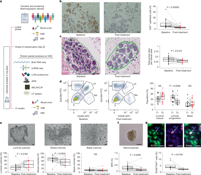
The study employed Taqman universal PCR master mix (Applied biosystems, 4304437) with Thermo Fisher gene expression assays to measure the expression of TNFSF11 (Hs00243522_m1), SOX9 (Hs00165814_m1), and KIT (Hs00174029_m1), normalizing data against β-actin expression (Hs99999903_m1). Gene expression was assessed using a QuantStudio 5 Real-time PCR system (Thermo Fisher) and analyzed with QuantStudio Design and Analysis software (v2.6.0).
Protein levels were analyzed via SDS-PAGE and nitrocellulose membrane transfer, followed by incubation with primary antibodies – anti-SOX9 (sigma, AB5535), anti-KIT (R&D Biosystems, MAB332), and anti-β-actin (Sigma, A1978) – at dilutions of 1:1,000, 1:1,000, and 1:5,000 respectively. Secondary antibodies (Dako, goat anti-rabbit 41424306 and anti-mouse 41424131, both at 1:5,000 dilution) were used for detection with Classico, Forte (Millipore) or West Femto (Thermo Fisher) horseradish peroxidase reagents, visualized using a ChemiDoc Touch Imaging System (Bio-Rad). Densitometry was performed using Image Lab (v6.1; BioRad) with normalization to β-actin.
Statistical significance was persistent using the Wilcoxon test (via the ‘ggpubr’ package, v0.6.0) with *P* values less than 0.05 considered notable. Data are presented as median and interquartile range, connected by lines for paired data.
The clinical study received approval from the local ethics review committee and involved local researchers in all phases, from design to publication authorship. A detailed reporting summary is available as supplementary material.